Neonatology: Understanding Hyperbilirubinemia in Newborns
Explore the metabolic transition of bilirubin in newborns, the risk of hyperbilirubinemia, and the potential complications such as Kernicterus. Learn about clinical manifestations and essential considerations in neonatal care.

Download Presentation

Please find below an Image/Link to download the presentation.
The content on the website is provided AS IS for your information and personal use only. It may not be sold, licensed, or shared on other websites without obtaining consent from the author. If you encounter any issues during the download, it is possible that the publisher has removed the file from their server.
You are allowed to download the files provided on this website for personal or commercial use, subject to the condition that they are used lawfully. All files are the property of their respective owners.
The content on the website is provided AS IS for your information and personal use only. It may not be sold, licensed, or shared on other websites without obtaining consent from the author.
E N D
Presentation Transcript
AL-Mustaqbal University College of Pharmacy Hospital Training 5ndStage Pediateric part Edited by: Dr. teba jasim mohammed Hospital Training Committee In Pharmacy Department /AL- Mustaqbal University College 1
Age Group Terminology Premature Birth before 37 completed weeks gestation Neonate 0-4 weeks Infant 1month-1 year Toddler 1-3 years Child 4-12 years Adolescent 13-18 years Adult >18 years 2
A- Neonatology 1-Hyperbilirubinemia in the Newborn (Neonatal Jaundice) Background 1- During the neonatal period, metabolism of bilirubin is in transition from the fetal stage, during which the placenta is the principal route of elimination of the lipid-soluble, unconjugated bilirubin, to the adult stage, during which the water soluble conjugated form is excreted from hepatic cells into the biliary system and gastrointestinal tract. 2- Bilirubin is derived primarily from the breakdown of heme-protein in the reticuloendothelial system. Nonpolar and water-insoluble unconjugated bilirubin is conjugated inside liver cells by the enzyme glucuronyl transferase to form Water-soluble conjugated bilirubin 3- Most conjugated bilirubin is excreted through the bile into the small intestine and eliminated in the stool. Some bilirubin may undergo hydrolysis back to the unconjugated fraction by intestinal glucuronidase, and may be reabsorbed (enterohepatic recirculation) 4- Nearly all newborns develop transient hyperbilirubinemia (serum bilirubin >2 mg/dL) and nearly 65% (two third) are clinically jaundiced (serum bilirubin >3mg/dL) 3
History of The Patient 5- Kernicterus (Bilirubin Encephalopathy) results when indirect (unconjugated) bilirubin is deposited in brain cells and disrupts neuronal function 6- Onset of jaundice in the first 24 hours of life is always pathological 7- Kernicterus (Bilirubin Encephalopathy) results when indirect (unconjugated) bilirubin is deposited in brain cells and disrupts neuronal function. 8- Kernicterus usually does not develop in term infants when bilirubin levels are less than 20 to 25 mg/dL. The incidence of kernicterus increases as serum bilirubin levels increase to greater than 25 mg/dL Kernicterus may be noted at bilirubin levels less than 20 mg/dL in the presence of some conditions like sepsis, meningitis, and prematurity 4
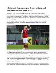
Clinical Manifestations Jaundice from deposition of indirect bilirubin in the skin tends to appear bright yellow or orange, while jaundice of the obstructive type (direct bilirubin) has a greenish or muddy yellow cast. Jaundice usually starts on the face and progress to the abdomen and then the feet, as serum levels increase. Yellow discoloration of the eyes usually occurs. Light-colored stools and dark urine are usually associated with pathologic hyperbilirubinemia. Infants with severe hyperbilirubinemia may present with lethargy and poor feeding and, without treatment, can progress to acute bilirubin encephalopathy (kernicterus). Symptoms and signs of kernicterus start with lethargy, poor feeding, and loss of the Moro reflex. Subsequently, the infant may appear gravely ill and prostrate, with diminished tendon reflexes and respiratory distress. Opisthotonos with a bulging fontanel, twitching of the face or limbs, and a shrill, high-pitched cry may follow. In advanced cases, convulsions and spasm occur, with affected infants stiffly extending their arms in an inward rotation with the fists clenched. Many of the neonates and infants who progress to these severe neurologic signs die, and the survivors are usually seriously damaged 5
Bulging fontanel Opisthotonos 6
Unconjugated hyperbilirubinemia 1-Nonpathologic unconjugated hyperbilirubinemia A-Physiologic Jaundice 1-Physiologic jaundice is an unconjugated hyperbilirubinemia that occurs after the first postnatal day and can last up to 1 week. Total serum bilirubin (TSB) concentrations peak in the first 3 to 5 postnatal days and decline to adult values over the next several weeks 2-The underlying mechanisms for physiologic jaundice in newborn are related to: (a) Increased bilirubin production because of elevated red blood cell volume per body weight and a shorter and shorter life span (b) Infants have immature hepatic glucuronyl transferase, a key enzyme involved in the conjugation of bilirubin (c) Increased enterohepatic circulation in newborn B- Breast milk jaundice 1-It occurs in some breast-fed infants because breast milk may contain an inhibitor of bilirubin conjugation or may increase the enterohepatic recirculation of bilirubin because of breast milk glucuronidase 2-Jaundice appears in the seventh day and it gradually increased in severity till it reaches its peak during third week It may persist for several weeks 3-Interruption of breast feeding and use of formula feeding for 1 3 days causes a prompt decline in bilirubin (which do not increase significantly after breastfeeding resumes) but is only recommended for infants with serumbilirubin concentrations that put them at risk for kernicterus
C-Breast feeding jaundice Breastfeeding jaundice occur when a breastfeeding baby is not getting enough breast milk, which leads to infrequent bowel movements and increased enterohepatic circulation of bilirubin. It occurs during the first week of life) Lower milk intake before breast milk production is established can result in dehydration, which hemoconcentrates bilirubin, while also causing fewer bowel movements, which in turn increases the enterohepatic circulation of bilirubin. Frequent breastfeeding (>10 in 24 hr), rooming-in with night feeding, and ongoing lactation support may reduce the incidence of early breastfeeding jaundice. In addition, supplementation with formula or expressed breast milk is appropriate if the intake seems inadequate, weight loss is excessive, or the infant appears dehydrated. D-Prematurity 1-Although preterm infants develop hyperbilirubinemia by the same mechanisms as term infants, it is more common and more severe in preterm infants and lasts longer (due to the relative immaturity of the red blood cells, hepatic cells, and gastrointestinal tract) 2-Kernicterus is extremely uncommon. However, kernicterus in preterm infantscan occur at lower TSB concentrations 8
2- Pathologic Unconjugated Hyperbilirubinemia A-Acute Hemolysis: In this condition, jaundice appears at birth or during the first day and it is commonly severe. Serum bilirubin level may rise rapidly to reach serious levels where kernicterus may occur. Kernicterus is a real risk and it may occur when serum bilirubin exceeds the critical level, which depends on the birth weight and the condition of the baby. The critical level is lower in those with low birth weight and in sick neonates The cause of haemolysis can be identified by clinical and laboratory evaluation. 1-Rh incompatibility: It is the commonest cause of hemolysis. It occurs in some Rh positive babies born to Rh negative mothers. Hemolysis occurs due to placental passage of maternal antibodies active against the fetal red cells. The first baby is usually not affected as maternal sensitization usually occurs during delivery of the first baby Rh incompatibility can be prevented by injection of Rh immune globulin to the mother within 72 hours after delivery which prevents her from forming antibodies which might affect subsequent babies 2-ABO incompatibility: ABO incompatibility may occur if the mother s blood type is O and the infant s blood type is A or B The first baby may be affected. Jaundice is not severe. kernicterus is rare. 9
B- Neonatal septicemia: 1-Jaundice in septicemia, if present, usually appears between the fourth and seventh day or later and is usually moderate in severity 2-The most important clinical signs are the markedly affected general condition The baby is not doing well with lethargy, poor suckling, fever or hypothermia, Immediate hospitalization and combined parenteral antibiotic therapy are important C-Other rare causes :(Conjugated Hyperbilirubinemia) 1-Conjugated (Direct-reacting) hyperbilirubinemia is never physiologic and should always be evaluated thoroughly 2-Direct-reacting bilirubin (composed mostly of conjugated bilirubin) is not neurotoxic to the infant, but signifies a serious underlying disorder involving hepatitis, cholestasis, hepatocellular injury or biliary atresia .atresia is an unusual closing or absence of a tube in the body). Biliary Atresia: Is an obstruction of the biliary tree that causes severe cholestasis and is characterized by elevation of the conjugated, or direct, bilirubin fraction , which leading to cirrhosis and death if left untreated in a timely manner . The jaundice of biliary atresia usually is not evident immediately at birth, but develops in the first week or two of life. The reason is that extrahepatic bile ducts are usually present at birth, but are then destroyed by an idiopathic inflammatory process 10
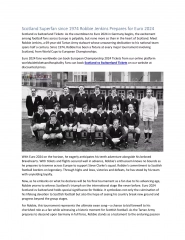
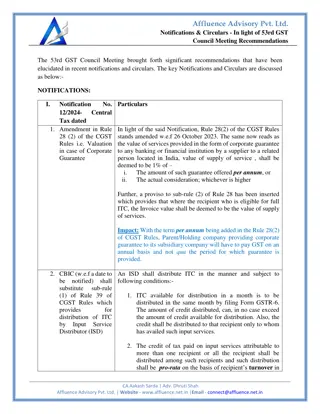
Therapy of Indirect (unconjugated) Hyperbilirubinemia The main concern is to prevent Kernicterus Charts exist indicating levels at which treatment should be initiated. Treatment options are: A- Phototherapy B- Exchange transfusion Phototherapy Healthy term baby Exchangetransfusion Healthy babyterm Preterm or any risk factors* Preterm or any risk factors Mg/dl mol/l Mg/dl Mg/dl Mg/dl mol/l mol/l m ol/l Day1 Day2 Day3 Day 4 and after Anyvisible jaundice** 15 18 20 15 260 425 510 510 13 15 20 20 220 260 340 340 260 310 340 13 16 17 22025 27030 29030 * Risk factors include small size ( less than 2.5 kg or born before 37 weeks gestation), haemolysis, and sepsis. ** Visible jaundice anywhere on body on day 1. 11
A-Phototherapy 1-Bilirubin absorbs light maximally in the blue range (420-470 nm), so blue light (not ultraviolet) of wavelength 450 nm is used to convert the bilirubin in the skin and superficial capillaries into harmless water-soluble metabolites, which are excreted in urine and through the bowel . 2-The eyes are covered to prevent discomfort and additional fluids are given to counteract increased losses from skin . 3-Serum bilirubin monitoring should continue for at least 24 hr after cessation of phototherapy in patients with hemolytic disease, because unexpected rises in bilirubin may occur, requiring further treatment . B-Exchange transfusion 1-This is required if the bilirubin rises to levels considered dangerous despite phototherapy . 2-Twice the infant's blood volume (i.e. 2 x 80 mL/kg) is exchanged over about 2 hours (or 2 x 85 mL/kg) . 3-The procedure is carried out through umbilical vein catheter . 4-Potential complications from exchange transfusion include metabolic acidosis, electrolyte abnormalities, hypoglycemia, hypocalcemia, thrombocytopenia, volume overload, arrhythmias, NEC, infection, graft-versus- host disease, and death. 12
C- Pharmacological agents 1-High dose intravenous immunoglobulin (IVIG) (0.5-1.0 g/kg/dose; repeat in 12 hr) is used in clinical practice for infants presenting with high jaundice levels secondary to rhesus or ABO incompatibility, it reduces the need for exchange transfusion presumably by reducing hemolysis . 2-Metalloporphyrins is a possible adjunct therapy for hyperbilirubinemia. The metalloporphyrin Sn mesoporphyrin (SnMP) offers promise as a drug candidate. A single intramuscular dose on the 1st day of life may reduce the need for subsequent phototherapy. Such therapy may be beneficial when jaundice is anticipated, particularly in patients with Rh incompatibility, ABO incompatibility or G6PD deficiency . Note: Water and dextrose solutions should not be used to supplement breastfeeding because they do not prevent hyperbilirubinemia and may lead to hyponatremia secondary to free water retention, which can lead to some serious signs and symptoms including nausea, vomiting, apathy, headache, seizures, hypothermia, weakness, and coma . 13
Management of conjugated hyperbilirubinemia Management depend on the treatment of the causative diseases (e.g. biliary atresia) . Treatment of extrahepatic biliary atresia is the surgical Kasai procedure, in which the fibrotic extrahepatic bile duct remnant is removed and replaced with a roux-en-Y loop of jejunum. This operation must be performed before 3 months of age to have the best chance of success . many children require liver transplantation . 2-Neonatal Sepsis and Meningitis 1- Neonates, especially preterm newborns, are at increased risk for infections and should be considered immunocompromised . 2- Risk factors of neonatal sepsis include prematurity, low birth weight, and predisposing maternal conditions (e.g., urinary tract infection) . 3- Early-onset neonatal sepsis (sepsis that presents during the first 7 days of life) usually is caused by organisms acquired from the maternal genital tract. (see table 1) 4- Late-onset sepsis (8 to 28 days) usually occurs in a healthy full-term infant who was discharged in good health. (see table 1) 5- Meningitis occurs as a complication of bacterial sepsis . The major pathogens causing neonatal sepsis are also the primary pathogens that cause neonatal meningitis . 14
table 1 15
Clinical Manifestations of Neonatal Sepsis 1-The most common signs are poor feeding, temperature instability (Hypothermia is more common than fever in neonatal sepsis, especially in preterm newborns), lethargy, or apnea . 2-Other signs of neonatal sepsis include tachycardia, dyspnea or cyanosis, tachypnea, disseminated intravascular coagulation (DIC)and abdominal distension . 3-The clinical manifestations of sepsis are difficult to separate from the manifestations of meningitis in the neonate. Laboratory Diagnosis of Neonatal Sepsis A- Positive cultures of body fluids confirm the diagnosis, including the following: 1. Blood: Must be obtained as a part of every evaluation for sepsis. 2. CSF: CSF analysis is indicated for all infants with a positive blood culture. 3. Urine: urine cultures are indicated, as urinary tract infections are a frequent source of infection . B- Hematologic studies: 1.An extremely elevated total WBC or very depressed count is more suggestive of infection. 2.Thrombocytopenia is also associated with sepsis . C-A chest radiograph is indicated in all infants with respiratory symptoms . D-C-reactive protein(CRP): CRP levels are often elevated in neonatal patients with bacterial sepsis E-Coagulation studies : prolonged values may indicate DIC 16
Treatment of Sepsis and Meningitis 1-The initial empiric antibiotic treatment of choice for early-onset neonatal sepsis and meningitis is ampicillin plus an aminoglycoside (Tables 2 and 3) . [In some nurseries, a third- generation cephalosporin (e.g., cefotaxime), instead of an aminoglycoside is added to ampicillin]. 2-If meningitis is highly suspected, gentamicin may be replaced by a third generation cephalosporin(cefotaxime) owing to greater CSF penetration . 3-For late-onset sepsis or meningitis , a combination of vancomycin with an aminoglycoside (gentamicin or tobramycin) is appropriate . 4-Amphotericin remains the treatment of choice for invasive candidiasis when meningitis is a consideration; liposomal amphotericin or anechinocandin (caspofungin or micafungin) are options for hepatic or splenic candidiasis. Fluconazole might be an effective therapy for susceptible organisms Duration of therapy 1-Therapy for most bloodstream infections should be continued for a total of 7- 10 days or for at least 5-7 days after a clinical response has occurred . 2-Meningitis should be treated for 14-21 days . 17
Table 3 t Table 1 18
Supportive care 1- Fluids, electrolytes, and glucose levels should be monitored carefully with correction when needed . 2- Seizures should be treated with anticonvulsants . 3- DIC may complicate neonatal septicemia. DIC may require fresh frozen plasma, platelet transfusions, or whole blood. 4- The use of intravenous immunoglobulin (IVIG) has been shown to decrease mortality in patients with sepsis . 19
B-Nephrology 1-Nephrotic syndrome (NS) is characterized by persistent heavy proteinuria (mainly albuminuria) ; hypoproteinemia (serum albumin <3.0 g/dL); hypercholesterolemia (>250 mg/dL); and edema . 2-Nephrotic syndrome is primarily a pediatric disorder and is 15 times more common in children than adults (2) with a peak age of onset in children aged<6yrs . 3-The underlying abnormality in nephrotic syndrome is an increase in permeability of the glomerular capillary wall, which leads to massive proteinuria and hypoalbuminemia . 4-Hypoalbuminemia causes a decrease in the plasma oncotic pressure and shift of fluid from the intravascular compartment to the interstitial space. Reduced plasma volume stimulates antidiuretic hormone (ADH) secretion and the renin angiotensin system, producing sodium and water retention, exacerbating the edema . 20
Classification Approximately 90% of children with NS have idiopathic NS. Idiopathic NS includes three histologic types : A-Minimal change nephrotic syndrome (MCNS) is the most common form of NS in children (accounts for about 85%) B-Other less common types are [Focal segmental glomerulosclerosis (FSGS), and Membranoproliferative glomerulonephritis (MPGN) ] . Clinical features 1-Children usually present with mild edema, which is initially noted around the eyes (Periorbital ) and in the lower extremities . Periorbital oedema is often most noticeable in morning on rising . 2-With time, the edema becomes generalized, with the development of ascites, pleural effusions, and genital edema . Treatment 1-NS edema is treated by restricting salt intake. Severe edema may require the use of loop diuretics. When these therapies do not alleviate severe edema, parenteral administration of 25% albumin (0.5 to 1.0 g/kg intravenously over 1 to 2 hours) with an intravenous loop diuretic usually results in diuresis . 2-Children with onset of nephrotic syndrome between 1 and 8 yr of age are likely to have steroid-responsive minimal change disease, therefore, steroid therapy (prednisolone 2 mg/kg/day)( 60 mg/m2/day) may be initiated without renal biopsy . 21
Treatment 3-After the initial 4-6 wk course, the prednisone dose should be tapered to 40 mg/m2/day given every other day as a single morning dose. The alternate-day dose is then slowly tapered and discontinued over the next 2-3 mo . 4-Steroid-dependent patients (relapse while on alternate-day steroid therapy or within 28 days of stopping prednisone therapy), frequent relapsers, and steroid- resistant patients may be candidates for alternative agents (e.g. Cyclophosphamide). 5-Rituximab has been effective in the treatment of refractory NS in children, and it could reduce the use of steroid and immunosuppressants. 6-Acute hypertension (HTN) is treated with -blockers or calcium channel blockers. Persistent HTN usually responds to ACE inhibitors . 7-ACE inhibitors and angiotensin II blockers may be helpful as an adjunct therapy to reduce proteinuria in steroid-resistant patients. 22
Complication 1-Infection is the major complication of nephrotic syndrome. Children in relapse have increased susceptibility to bacterial infections owing to urinary losses of immunoglobulins and use of immunosuppressive therapy. Spontaneous bacterial peritonitis is the most frequent type of infection. 2-The role of prophylactic antibiotic therapy during relapse remains controversial. 3- Children with nephrotic syndrome are also at increased risk for Thromboembolism (TE). (related to increased prothrombotic factors (e.g. fibrinogen,) and decreased fibrinolytic factors) . Prophylactic anticoagulation is not recommended in children unless they have had a previous TE. Warfarin, low-dose aspirin, or dipyridamole may minimize the risk of clots in NS patients with a history of TE or high risk for TE Prognosis 1-The majority of children with steroid-responsive NS have repeated relapses, which generally decrease in frequency as the child grows older . 2-Steroid-responsive patients have little risk of chronic renal failure. Children with steroid-resistant NS, most often caused by focal segmental glomerulosclerosis, generally have a much poorer prognosis. These children develop progressive renal insufficiency, ultimately leading to end-stage renal failure requiring dialysis or renal transplantation 23
2-Hemolytic-Uremic Syndrome 1-The hemolytic-uremic syndrome (HUS) is the most common cause of acute renal failure in young children and is characterized by hemolytic anemia, thrombocytopenia, and uremia . HUS typically occurs in children less than 5 years of age but can occur in older children. 3-Two forms of HUS are recognized. A- HUS following infection with Shiga toxin-producing Escherichia coli (STEC-HUS or typical HUS, formally D+HUS) is the most common cause of HUS, responsible for up to 90% of cases in children. B-Atypical HUS (aHUS, formally D HUS) describes HUS in the absence of evidence of STEC infection. Note: D+ HUS diarrhea associated, D-HUS not diarrhea associated. 24
1- Classic D+HUS begins with gastroenteritis characterized by fever, vomiting, and diarrhea that is often bloody. Followed in 7 to 10 days by weakness, lethargy, and oliguria/anuria. Physical examination reveals irritability,pallor, and petechiae . Treatment and Prognosis 1-Therapy for HUS is supportive and includes volume repletion, and managing complications of renal insufficiency, including dialysis when indicated . 2- Red blood cell transfusions are provided as needed . 3-Antibiotics and antidiarrheal agents may increase the risk of developing HUS . [Antibiotics should be avoided in patients with acute enteritis presumed secondary to E. coli as they may increase the risk of developing HUS. 4- Most children (>95%) with D+HUS survive the acute phase and recover normal renal function, although some may have evidence of long-term morbidity. 25
C- Infection 1- Bronchiolitis: a lower respiratory tract infection (LRTI) that primarily affects the small airways (bronchioles), is a common cause of illness and hospitalization in infants and young children. 2-Bronchiolitis is seasonal, with peak activity during winter and early spring. 3-Bronchiolitis occurs almost exclusively during the first 2 years of life, with a peak age at 2 to 6 months . 4-Acute bronchiolitis is characterized by bronchiolar obstruction with edema, mucus, and cellular debris Etiology 1-Acute bronchiolitis is predominantly a viral disease. Respiratory syncytial virus (RSV) is responsible for more than 50% of cases . 2- Other agents include parainfluenza, adenovirus, Mycoplasma, and occasionally other viruses . 26
Clinical Manifestations. 1-The infant first develops a mild upper respiratory tract infection with sneezing and clear rhinorrhea. This may be accompanied by diminished appetite and fever . 2-Gradually, respiratory distress ensues, with paroxysmal wheezy cough, dyspnea, and irritability. The infant is often tachypneic, which interferes with feeding . 3-As a result of limited oral intake due to coughing combined with fever, infants are frequently dehydrated . Diagnosis The diagnosis of bronchiolitis is based primarily on history and clinical findings (6). Treatment 1-The mainstay of treatment is supportive. Therapy of bronchiolitis primarily consists of administration of supplemental oxygen and replacement of fluid deficits (hydration) as needed . 27
2-The risk of aspiration of oral feedings may be high in infants with bronchiolitis owing to tachypnea and the increased work of breathing. The infant may be fed through a nasogastric tube. 3-A number of agents have been proposed as adjunctive therapies for bronchiolitis: A- Bronchodilators produce modest short-term improvement in clinical features. Nebulized epinephrine may be more effective than -agonists . B- Corticosteroids, whether parenteral, oral, or inhaled, are widely used despite conflicting studies. C- Ribavirin, is a compound with antiviral activity against RSV administered by aerosol, has been used for infants with congenital heart disease (CHD)or chronic lung disease (CLD) although its benefit is uncertain . D-Antibiotics have no value unless there is secondary bacterial pneumonia 28
Prophylaxis 1-Palivizumab is a monoclonal antibody to RSV and can be used as prophylaxis initiated just before the onset of the RSV season (monthly IM injection for 5months starting in October) confers some protection from severe RSV disease. 2- Palivizumab is indicated for some infants under 2 years old with CLD, severe CHD or prematurity 29


 Hyperbilirubinemia")