Neoplasms of the Maxillofacial Area: Understanding Tumors and Oncology
This lecture delves into the definition of tumors, general aspects of oncology, and the classification of tumors in the maxillofacial area. It discusses modern diagnostic methods and the incidence of neoplasms in the face and jaws. Various perspectives on tumors are explored, providing a comprehensive overview of tumor growth and development.

Download Presentation

Please find below an Image/Link to download the presentation.
The content on the website is provided AS IS for your information and personal use only. It may not be sold, licensed, or shared on other websites without obtaining consent from the author.If you encounter any issues during the download, it is possible that the publisher has removed the file from their server.
You are allowed to download the files provided on this website for personal or commercial use, subject to the condition that they are used lawfully. All files are the property of their respective owners.
The content on the website is provided AS IS for your information and personal use only. It may not be sold, licensed, or shared on other websites without obtaining consent from the author.
E N D
Presentation Transcript
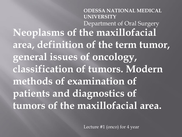
ODESSA NATIONAL MEDICAL UNIVERSITY Department of Oral Surgery Neoplasms of the maxillofacial area, definition of the term tumor, general issues of oncology, classification of tumors. Modern methods of examination of patients and diagnostics of tumors of the maxillofacial area. Lecture #1 (onco) for 4 year
The purpose of the lecture: To introduce statistic indices, incidence of neoplasms of the maxillofacial area. To analyze the concept tumor and main trends in study of these processes; to introduce accessible classifications of neoplasms of the face and jaws and modern possibilities of their diagnostics.
Oncology is the science studying origin, development, incidence of tumors, possibilities of their diagnostics, treatment and prevention. Its subdivision is oncologic dentistry which studies all these problems in relation to the tumors located within the maxillofacial area.
Before analyzing in detail certain types of tumors, it is necessary to understand what a tumor is, i.e. what is meant by this term. There are many definitions based on the viewpoint that tumor is an autonomic process in tissues connected with structural and functional changes on the cellular and subcellular levels.
One of the founders of domestic oncology A.A. Petrov (1961) regarded tumor as dystrophic proliferative response of the organism to an impact of irritating internal or external factors, which steadily affects the structure and contents of cells and tissues altering their metabolism . More complicated definition was given by the greatest pathologist I.V. Davydovsky who considered that cambial or normal tissues, when a certain stimulus is applied, acquire ability to irregular proliferation, but the factor of differentiation and development of cells disappears.
Lviv oncologist Gnatyshak (1988) defines tumor as specific form of tissue growth possessing a certain autonomy . Finally the most common current definition regards tumor as an abnormal mass of tissue with excessive growth which is not coordinated with growth of normal tissue and proceeds the same excessive way after the causative agent stops acting . In addition to these definitions, a number of others can be given (Yu.I. Bernadsky, A.A. Timofeev etc).
As can be seen from the aforementioned definitions, they classify already formed process without clarifying its causes. Many aspects of oncogenesis still continue to be unclear and poorly studied, including those in dentistry. Enrichment of our knowledge will correct and add, probably even change the views of this complex biologic process.
Talking about the ways of solving the problem, three correlated trends can be mentioned: 1. Biologic, i.e. experimental oncology studying causes, regularities and mechanisms of tumor growth. 2. Individual, i.e. study of causes of origin, pathogenesis and clinical signs of developing tumor in a person, development of methods of diagnostics, treatment and prevention of particular manifestations of tumors of various location and genesis in a certain person clinical oncology. 3. Social, i.e. study of incidence (epidemiology) of tumors, conditions of their origin and development; gender and age structure of the patients etc.
Summary and generalization of the data obtained in each trend allow to improve results of treating a particular patient and develop preventive measures for oncologic processes. Special epidemiologic investigations concerning incidence of tumors of the maxillofacial area are few. Moreover, as will be shown below, they are very diversified including benign and malignant tumors, tumor-like diseases with irregular structure.
Malignant tumors are characterized by atypical cells, incomplete process of their formation, invasive growth, ability to metastaze, i.e. transport tumor cells with blood flow, flow of lymph, tissue liquid to remote from the primary location zones (metastasis from Greek halted in another place ), quick spread in tissues.
Benign tumor retains cellular structure typical for the tissue from which it is formed, as a rule, has a clear capsule separating it from surrounding tissues; it is characterized by slow growth pushing aside surrounding tissues.
Malignant tumors of organs of the oral cavity and face rank the 7th place (4.5%) among tumors in man. Besides, they are 6-7 times more prevalent in men than in women. With advancing age their number dramatically increases: from 0.4/100,000 at the age of 30 to 69-75/100,000 in people over 60. An average index in Ukraine ranges from 3.9/100,000 in women to 22.6/100,000 in men.
The most common classification for today is the classification introduced by the expert committee of the WHO (1974, 1980): International Histologic Classification of Tumors including among others tumors of the maxillofacial location. Based upon this, narrower classifications using organo-morphologic characteristics (A.A. Kolesov, I.I. Ermolaev, V.P.Panikarovsky, A.M. Solntsev etc) have been introduced. Histologic characteristic depends on the origin of the tissue: epithelial, muscular, osseous, connective etc. Frequently tumor contains several histologic components which also complicates diagnostics.
The tumor can be classified only as a result of thorough clinical investigation, additional methods (radiography, ultrasound etc) and morphologic investigation. Let us analyze these possibilities in details.
Clinical examination includes thorough anamnesis, careful examination of tissues and organs of the oral cavity, palpation. Today several levels of diagnostics are distinguished: extra-early based mainly on the genetic level, family history, congenital and inherited diseases. This diagnostics is virtually never used in dentistry. Preclinical or early clinical diagnostics is based on determination of risk groups, knowledge of epidemiology of some diseases, life conditions, work conditions, detected precancerous diseases.
Modern diagnostics allows to detect tumor processes at early stages. During clinical examination the following factors should be considered: syndrome of plus tissue or erosion , presence of pathologic discharges, pains of unclear character, impaired functions of salivation, mimic muscles, tongue movements, nasal respiration etc. The analysis and results of this examination can be summarized in the following statement: ability to diagnose any pathologic condition is not a mandatory requirement of a medical specialist, but ability to promptly suspect any deviation from normal and subsequently direct a patient to a specialist is a must. Detecting a tumor at early stages provides, as a rule, its complete cure. At later stages survivability can amount to under 10%.
The most available, depending on location and character of a tumor is cytologic investigation of the swabs, prints, scrapes to determine the character and type of cellular elements. Stomatoscopy should be widely used with vital staining of suspected structures and surrounding tissues, especially, in affected oral mucosa. Other non- invasive methods are radioisotope and ultrasound diagnostics, thermovisiography which sometimes (scanning) allow to verify the character of the tumor but more frequently simply detect impaired growth or structure of the investigated tissue.
Common methods include various types of radiography from studying radiographs taken in a standard position to application of CT, MRI allowing to detect changes not only in osseous but in soft-tissue structures as well. For investigation of cavities and glandular formations more complete information can be provided by contrast methods of radiographic investigation.
The most reliable information can be obtained with morphologic investigation of the tissue region taken by means of biopsy: fine needle aspiration biopsy, incision biopsy, excision biopsy. To take the material, a number of technologic requirements should be strictly followed, which determine possibility of the investigation. For some types of tumors immune diagnostics is used, level of oncogenes, size and types of lymphocytes, level and structure of a number of aminoacids determined. These methods do not have much value in diagnosing tumors in the maxillofacial surgery.
Thus, the incidence of oncologic lesions of the maxillofacial area is quite high, and detection of advance staged tumors witnesses insufficient qualification of doctors of various levels. That is why possibilities of diagnostics should be used by every doctor.


 defines tumor as")