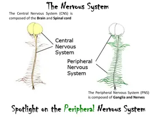
Neural Activity and Function in the Nervous System
Explore the effects of wheel-running activity on the integration of photoperiodic changes in the suprachiasmatic nucleus, static markers of neural activity, and methods for visualizing neural activity such as protein trafficking, voltage-sensitive dye imaging, synaptic transmission, and calcium dynamics.

Download Presentation

Please find below an Image/Link to download the presentation.
The content on the website is provided AS IS for your information and personal use only. It may not be sold, licensed, or shared on other websites without obtaining consent from the author. If you encounter any issues during the download, it is possible that the publisher has removed the file from their server.
You are allowed to download the files provided on this website for personal or commercial use, subject to the condition that they are used lawfully. All files are the property of their respective owners.
The content on the website is provided AS IS for your information and personal use only. It may not be sold, licensed, or shared on other websites without obtaining consent from the author.
E N D
Presentation Transcript
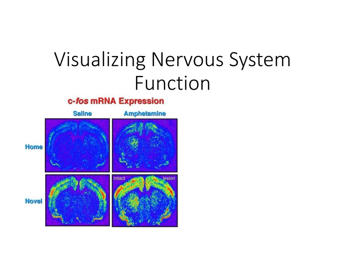
Visualizing Nervous System Function
Effects of wheel-running activity on the integration of a photoperiodic change by the suprachiasmatic nucleus (SCN). Hamsters were exposed to the short photoperiod [10:14-h light-dark cycle (LD10:14)] for only 1 day (LPLP groups), for 4 wk (LPSP groups), or for 12 wk (SPSP groups). Some hamsters had free access to a running wheel for 4 wk before death, and some hamsters were light stimulated at the end of the night. See materials and methods for details. A: representative photomicrographs of c-Fos immunoreactivity within the SCN. Scale bar = 250 m. B: quantification of c-Fos immunoreactivity (ir) within the SCN of hamsters exposed to different light-dark conditions, with or without a wheel, and light stimulated or not (n = 5 in each group). Values are mean SE.
Static Markers of Activity Assaying Neural Activity in Fixed Tissue Immediate Early Genes (IEGs) Encode diverse range of proteins, not often used as a stand-alone indicator of activity Assaying Cellular Function in Fixed Tissue Cell proliferation Thymidine Analogs BrdU (bromo-deoxyuridine), a synthetic analog of the DNA base thymidine, or radioactive tritiated thymidine (3H-thymidine) KI67 - During interphase, the antigen can be exclusively detected within the nucleus, whereas in mitosis most of the protein is relocated to the surface of the chromosomes. The fact that the Ki-67 protein is present during all active phases of the cell cycle (G(1), S, G(2), and mitosis), but is absent from resting cells (G(0)), makes it an excellent marker for determining the so-called growth fraction of a given cell population
Visualizing Neural Activity Protein Trafficking Pulse-chase Labeling Imaging Voltage Voltage-Sensitive Dye Imaging (VSDI) Primary method to visualize changes in voltage These dyes shift their absorption based on membrane potential Able to detect subthreshold synaptic potentials Imaging Synaptic Transmission FM dyes Lipophilic styryl dyes that fluoresce when bound to membranes. Useful for exocytosis and endocytosis Neurotransmitters Release examined using genetically encoded synaptic transmission reporter synapto-pHluorin, which uses pH- sensitive mutants of GFP
Visualizing Neural Activity Imaging Calcium Calcium central to many physiological processes Calcium dynamics link electrical activity and biochemical events Indicator dyes are organic molecules that change their spectral properties when bound to Ca 2+
Optically Manipulating Neural Activity Stimulation through Uncaging Molecules https://www.youtube.com/watch?v=MWCoz_ghNtQ Light-Activated Channels SPARK (synthetic photoisomerizable azobenzene- regulated K+) channels are potassium channels fused to a molecular tether containing an ion that can block the channel pore. Long wavelengths (500nm green) block the channel pore, short wavelengths (380nm blue) allow efflux of K ions out. Using light instead of electrical stimulation is beneficial because it eliminates artifacts.
Visualizing Protein Function Time-Lapse Imaging with Reporter Genes Non-endogenous gene encoding an enzyme or fluorescent protein whose expression is controlled by a promoter for a separate gene of interest Fluorescence Resonance Energy Transfer (FRET) Used to monitor interactions between proteins Two molecules of interest labeled with different fluorophore, each chosen such that the emission spectrum of one overlaps with the absorption of the other
Visualizing Protein Function Biomolecular Fluorescence Complementation (BiFC) Similar to FRET, but rather than two separate fluorophores, two halves of a single fluorophore is fused to one protein and does not fluoresce. When two halves are close enough, they fold and form a single functional fluorophore Fluorescence Recovery after Photobleaching (FRAP) Limitation of fluorophores is that photobleaching results in decreased fluorescence intensity over time This technique uses strong laser illumination to bleach the fluorescent molecules in a particular region Then measures the time-course of fluorescence returning to the bleached region
Visualizing Protein Function Photoactivation & Photoconversion Photoactivation When some fluorescent proteins only activate when hit with a specific wavelength, called Photoactivation Photoconversion Serves a similar purpose, but rather than activating a previously non-fluorescent molecule, a pulse of light causes a fluorophore to change its emission spectra from one color to another
Optically Manipulating Protein Function Photo-activation/ Photo-Uncaging Technique can be used more generally for optical control over many molecules Photo-inactivation Chromophore-assisted laser inactivation (CALI) is a method used to inhibit the activity of specific proteins in precise subcellular regions
Visualizing Nervous System Function Summary Reporter Genes Useful for observing behaviour, localization, or movements of tagged protein FRET Useful for observing interactions between two tagged proteins or two parts of a single protein BiFC Measures change in fluorescent intensity to indicate whether the two proteins are near each other FRAP Useful for observing dynamics of trafficking, diffusion, or binding/dissociation of fluorescently tagged proteins Photoactivation/Photoconversion Useful for tracking localization changes of small populations or individual cells, organelles, or proteins.
Visualizing Nervous System Function Summary Photoactivation/Photo-Uncaging Light removes an inhibiting chemical group from a caged compound. Rapid acting with experimenter control over spatial and temporal release of compounds CALI Light stimulates chromophore to produce ROS that destroy targeted proteins. Proteins can be targeted through the use of antibodies or genetic tags