Neurological Stress and Epigenetics in Physical Rehabilitation: Recent Advances
Neurological stress can have profound effects on the body, particularly through the activation of the HPA axis. This presentation delves into the intricate relationship between stress, epigenetics, and physical rehabilitation, exploring recent research on mental flexibility deficits in PTSD and mTBI, glucocorticoid-related predictors of PTSD treatment success in combat veterans, and the impact of different types of exercise on DNA methylation patterns.

Download Presentation

Please find below an Image/Link to download the presentation.
The content on the website is provided AS IS for your information and personal use only. It may not be sold, licensed, or shared on other websites without obtaining consent from the author.If you encounter any issues during the download, it is possible that the publisher has removed the file from their server.
You are allowed to download the files provided on this website for personal or commercial use, subject to the condition that they are used lawfully. All files are the property of their respective owners.
The content on the website is provided AS IS for your information and personal use only. It may not be sold, licensed, or shared on other websites without obtaining consent from the author.
E N D
Presentation Transcript
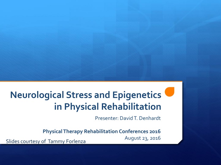
Neurological Stress and Epigenetics in Physical Rehabilitation Presenter: David T. Denhardt Physical Therapy Rehabilitation Conferences 2016 August 23, 2016 Slides courtesy of Tammy Forlenza
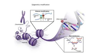
Neurological Stress and Epigenetics in Physical Rehabilitation OUTLIINE: (No major role for OPN) (REVIEW OF SOME RECENT IMPORTANT ADVANCES NOT MY OWN) David T. Denhardt, Rutgers University, Emeritus Professor denhardt@dls.rutgers.edu Aug. 23, 2016 Neurological Stress The cause of multiple problems in the world today many consequences of varying degrees of severity results in the activation of the HPA axis [hypothalamus (vasopressin and corticotropin releasing hormone, CRH), pituitary (adrenal corticotropic hormone, ACTH), and adrenals (cortisol, CORT)] glucocorticoid & mineralocorticoid release - essential roles in signaling most of the stress responses thought out the body engage a variety of receptors in numerous target cells throughout the body and brain. Presented following this intro are details of 5 papers. E.W. Pang and colleagues: (Univ of Toronto, Canada) Different neural mechanisms underline deficits in mental flexibility . (Frontiers Psychiatry 2015) Mental inflexibility core executive function similar in both PTSD (post-traumatic stress disorder) and mTBI (mild traumatic brain injury). Distinct neural profiles in the two disorders evidenced by magnetoencephalography: Reduced brain connectivity and mental flexibility in mTBI (Clin Translational Neurol2015) shown by MEG. When challenged, patients unable to boost their connectivity, hence a deterioration in performance. Limits access to one s cognitive reserve: From Structure to Circuits: The Contribution of MEG Connectivity Studies to Functional Neurosurgery (Frontiers in Neuroanatomy 2016) Based on the fact that intracellular neuronal currents generate a magnetic field that can be detected by highly sensitive biomagnetometers. Offers exquisite resolution in time and oscillatory domain in a spatial domain. Enhances what is seen with fMRI connectivity studies. R. Yehuda and co-workers: (VA Med Cntr, Icahn Sch of Med, Mt Sinai NY) Glucocorticoid-related predictors and correlates of PTSD treatment in combat veterans (Interface Focus, Royal Society 2014). The NR3C1 glucocorticoid receptor, notably the BCLI polymorphism therein, is associated with increased GR sensitivity, particularly the GG and GC alleles, appeared to be most predictive of successful recovery from PTSD. S. Horsburgh and colleagues (Dept Applied Science, Northumbria Univ, UK) Circulating changes following damaging eccentric exercise do not significantly alter markers of DNA methylation in human peripheral blood mononuclear cells . (Submitted to Frontiers in Genetics: Epigenomics and Epigenetics 2016). Aerobic exercise involves endurance training, which involves extensive time over repeated periods. Eccentric (jump training) requires muscles to exert maximum force in short intervals of time, with the goal of increasing power. Aerobic, but not eccentric, exercise leads to increased oxygen utilization and attenuation of global and gene-specific methylation. Eccentric ( drop jump ) leads to an increase in muscle soreness and plasma creatine kinase activity but no change in DNA methylation patterns. More broadly, exercise of either sort clearly has the potential, with some evidence, that that significant changes in systemic inflammation, likely via PBMCs that circulate throughout the body, may cause multiple epigenetic effects, likely by affecting miRNAs and multiple enzymes involved in modifying DNA.
Different Neural Mechanisms Underlie Deficits in Mental Flexibility in Post-Traumatic Stress Disorder Compared to Mild Traumatic Brain Injury Front Psychiatry. 2015 Dec 3;6:170 . E.W. Pang and colleagues: (University of Toronto, Ont. Canada) ABSTRACT: Mental flexibility is a core executive function that underlies the ability to adapt to changing situations and respond to new information. Individuals with post-traumatic stress disorder (PTSD) and mild traumatic brain injury (mTBI) complain of a number of executive function difficulties, one of which is mental inflexibility or an inability to switch between concepts. While the behavioral presentation of mental inflexibility is similar in those with PTSD or mTBI, we hypothesized that the differences in their etiology would manifest as differences in their underlying brain processing. The neural substrates of mental flexibility have been examined with a number of neuroimaging modalities. Functional magnetic resonance imaging has elucidated the brain regions involved, whereas electroencephalography has been applied to understand the timing of the brain activations. Magnetoencephalography, with its high temporal and spatial resolution, has more recently been used to delineate the spatiotemporal progression of brain processes involved in mental flexibility and has been applied to the study of clinical populations. In a number of separate studies, our group has compared the source localization and brain connectivity during a mental flexibility set-shifting task in a group of soldiers with PTSD and civilians with an acute mTBI. In this article, we review the results from these studies and integrate the data between groups to compare and contrast differences in behavioral, neural, and connectivity findings. We show that the different etiologies of PTSD and mTBI are expressed as distinct neural profiles for mental flexibility that differentiate the groups despite their similar clinical presentations.
Reduced brain connectivity and mental flexibility in mild traumatic brain injury Ann Clin Transl Neurol. 2015; 3:124-31 E.W. Pang and colleagues: (University of Toronto, Ont., Canada) OBJECTIVE: A mild traumatic brain injury (mTBI), or concussion, has known neuropsychological sequelae, and neuroimaging shows disturbed brain connectivity during the resting state. We hypothesized that task-based functional connectivity measures, using magnetoencephalography (MEG), would better link the neurobiological underpinnings of cognitive deficits to specific brain damage. METHODS: We used a mental flexibility task in the MEG and compared brain connectivity between adults with and without mTBI. RESULTS: Affected individuals showed significant reductions in connectivity. When challenged with a more difficult task, these individuals were not able to "boost" their connectivity, and as such, showed deterioration in performance. INTERPRETATION: We discuss these findings in the context of limitations in cognitive reserve as a consequence of a mTBI.
From Structure to Circuits: The Contribution of MEG Connectivity Studies to Functional Neurosurgery Front Neuroanatomyt2016 10:67 E .W. Pang and colleagues: (University of Toronto, Ont. Canada) ABSTRACT New advances in structural neuroimaging have revealed the intricate and extensive connections within the brain, data which have informed a number of ambitious projects such as the mapping of the human connectome. Elucidation of the structural connections of the brain, at both the macro and micro levels, promises new perspectives on brain structure and function that could translate into improved outcomes in functional neurosurgery. The understanding of neuronal structural connectivity afforded by these data now offers a vista on the brain, in both healthy and diseased states, that could not be seen with traditional neuroimaging. Concurrent with these developments in structural imaging, a complementary modality called magnetoencephalography (MEG) has been garnering great attention because it too holds promise for being able to shed light on the intricacies of functional brain connectivity. MEG is based upon the elemental principle of physics that an electrical current generates a magnetic field. Hence, MEG uses highly sensitive biomagnetometers to measure extracranial magnetic fields produced by intracellular neuronal currents. Put simply then, MEG is a measure of neurophysiological activity, which captures the magnetic fields generated by synchronized intraneuronal electrical activity. As such, MEG recordings offer exquisite resolution in the time and oscillatory domain and, as well, when co-registered with magnetic resonance imaging (MRI), offer excellent resolution in the spatial domain. Recent advances in MEG computational and graph theoretical methods have led to studies of connectivity in the time-frequency domain. As such, MEG can elucidate a neurophysiological-based functional circuitry that may enhance what is seen with MRI connectivity studies. In particular, MEG may offer additional insight not possible by MRI when used to study complex eloquent function, where the precise timing and coordination of brain areas is critical. This article will review the traditional use of MEG for functional neurosurgery, describe recent advances in MEG connectivity analyses, and consider the additional benefits that could be gained with the inclusion of MEG connectivity studies. Since MEG has been most widely applied to the study of epilepsy, we will frame this article within the context of epilepsy surgery and functional neurosurgery for epilepsy.
Glucocorticoid-related predictors and correlates of post- traumatic stress disorder treatment response in combat veterans Interface Focus. 2014 6;2014 R. Yehuda and co-workers: (VA Med Center, Icahn School of Med, Mt Sinai NY) ABSTRACT The identification of biomarkers for post-traumatic stress disorder (PTSD) and resilience/recovery is critical for advancing knowledge about pathophysiology and treatment in trauma-exposed persons. This study examined a series of glucocorticoid-related biomarkers prior to and in response to psychotherapy. Fifty-two male and female veterans with PTSD were randomized 2 : 1 to receive either prolonged exposure (PE) therapy or a weekly minimal attention (MA) intervention for 12 consecutive weeks. Psychological and biological assessments were obtained prior to and following treatment and after a 12-week naturalistic follow-up. Response was defined dichotomously as no longer meeting criteria for PTSD at post-treatment based on the Clinician Administered PTSD Scale for DSM-IV (CAPS). Clinical improvement on the CAPS was apparent for both PE and MA, with no significant difference according to treatment condition. Biomarkers predictive of treatment gains included the BCLI polymorphism of the glucocorticoid receptor gene. Additional predictors of treatment response were higher bedtime salivary cortisol and 24 h urinary cortisol excretion. Pre-treatment plasma dehydroepiandrosterone/cortisol ratio and neuropeptide Y (NPY) levels were predictors of reductions in PTSD symptoms, and, for NPY only, of other secondary outcomes as well, including anxiety and depression ratings. Glucocorticoid sensitivity changed in association with symptom change, reflecting clinical state. It is possible to distinguish prognostic and state biomarkers of PTSD using a longitudinal approach in the context of treatment. Identified markers may also be relevant to understanding mechanisms of action of symptom reduction.
Exercise and inflammation-related epigenetic modifications: focus on DNA methylation. Exerc Immunol Rev. 2015;21:26-41. S. Horsburgh and colleagues (Dept Applied Sci, Northumbria Univ, UK) ABSTRACT Epigenetics is the study of mitotically or meiotically heritable phenotypes that occur as a result of modifications to DNA, thereby regulating gene expression independently of changes in base sequence due to manipulation of the chromatin structure. These modifications occur through a variety of mechanisms, such as DNA methylation, post-translational histone modifications, and non-coding RNAs, and can cause transcriptional suppression or activation depending on the location within the gene. Environmental stimuli, such as diet and exercise, are thought to be able to regulate these mechanisms, with inflammation as a probable contributory factor. Research into these areas is still in its infancy however. This review will focus on DNA methylation in the context of inflammation (both pro- and anti-inflammatory processes) and exercise. The complexity and relative shortcomings of some existing techniques for studying epigenetics will be highlighted, and recommendations for future study approaches made.
Exercise-conditioned plasma attenuates nuclear concentrations of DNA methyltransferase 3B in human peripheral blood mononuclear cells. Physiol Rep. 2015 3. S. Horsburgh and colleagues (Dept Applied Science, Northumbria Univ, UK) ABSTRACT DNA methylation is modifiable by acute and chronic exercise. DNA methyltransferases (DNMT) catalyze this process; however, there is a lack of literature concerning the specific mechanisms by which exercise-induced modifications occur. Interleukin 6 (IL-6) stimulation of various cell lines has been shown to augment DNMT expression and nuclear translocation, which suggests a possible pathway by which exercise is able to elicit changes in epigenetic enzymes. The present study sought to elucidate the response of the de novo methyltransferases DNMT3A and DNMT3B to circulatory factors found in plasma isolated from whole blood before and after 120-min of treadmill running at an intensity of 60% of individual velocity at V O2max (vV O2max) interspersed with 30-sec sprints at 90% of vV O2max every 10- min. Peripheral blood mononuclear cells (PBMCs) isolated from a resting participant were incubated with plasma isolated from exercising participants (n =10) or recombinant IL-6 (rIL-6), followed by nuclear protein extraction and quantification of DNMT3A and DNMT3B concentrations. Nuclear concentrations of DNMT3B significantly decreased following the experimental protocol (P = 0.03), with no change observed in DNMT3A (P =0.514). Various concentrations of rIL-6 caused an elevation in both DNMT3A and DNMT3B nuclear concentration compared with the blank control. The conflicting results between exercising and rIL-6 conditions suggests that IL-6 does regulate DNMT nuclear transport, however, other plasma mediators may also exert significant influence on the nuclear concentrations of these enzymes.
Circulating Changes Following Damaging Eccentric Exercise Do Not Significantly Alter Markers of DNA Methylation in Human Peripheral Blood Mononuclear Cells S. Horsburgh and colleagues (Dept Applied Science, Northumbria Univ, UK) ABSTRACT Although research has begun to characterise epigenetic modifications associated with exercise, there is still a paucity of data regarding the mechanisms by which these changes occur. We have previously shown that exercise-conditioned plasma was able to attenuate nuclear concentrations of DNMT3B, whilst recombinant interleukin 6 augmented the nuclear concentrations of both DNMT3A and DNMT3B, suggesting differential regulation of the two enzymes. The present study, therefore, sought to elucidate the response of de novo DNMTs following a bout of eccentric exercise, and whether alterations in DNMT nuclear concentration exert a functional effect on global DNA methylation. 10 recreationally active males performed 100 drop jumps from a box measuring 0.6m, with assessment of subjective muscle soreness via a visual analogue scale, and muscle function via maximal voluntary contractions (MVC) of the quadriceps, immediately pre-, post-, 2h post-, 24h post-, 48h post-, and 72h post-exercise. Venous blood samples were taken at each time point, with exercise-conditioned plasma used to stimulate peripheral blood mononuclear cells (PBMCs) that had been isolated from a resting participant. Nuclear and cytoplasmic proteins were extracted, and DNMT3A/3B concentrations quantified using ELISAs. An ELISA based method was also used to quantify global DNA methylation. Muscle soreness increased immediately post-exercise and remained elevated at 72h post-exercise. There was a significant decrement in MVC, which returned to pre-exercise levels by 48h post-exercise. Plasma creatine kinase was augmented 2h post-exercise, peaking at 24h post- exercise. There was also a small, but statistically significant elevation in plasma IL-6 post-exercise. There were, however, no statistical changes in nuclear or cytoplasmic concentrations of DNMT3A or DNMT3B, nor global DNA methylation, at any time point. These data support the previous hypothesis that large, transient exercise-induced increases in plasma IL-6 associated with aerobic exercise could exert significant influence on cellular transport and/or transcriptional regulation of de novo DNMTs, in corroboration with previous data.