Explore a detailed analysis of early neurophysiological diagnosis techniques for Thoracic Outlet Syndrome (TOS). This study covers patient profiles, diagnostic methods, and results, providing valuable insights into the classification of TOS cases as unlikely, vascular, or neurogenic. The comparison with healthy subjects offers further perspective on nerve amplitude and latency criteria. Dive into the intricacies of TOS diagnosis and classification based on neurophysiological parameters and clinical features.
Please find below an Image/Link to download the presentation.
The content on the website is provided AS IS for your information and personal use only. It may not be sold, licensed, or shared on other websites without obtaining consent from the author. If you encounter any issues during the download, it is possible that the publisher has removed the file from their server.
You are allowed to download the files provided on this website for personal or commercial use, subject to the condition that they are used lawfully. All files are the property of their respective owners.
The content on the website is provided AS IS for your information and personal use only. It may not be sold, licensed, or shared on other websites without obtaining consent from the author.
E N D
Presentation Transcript
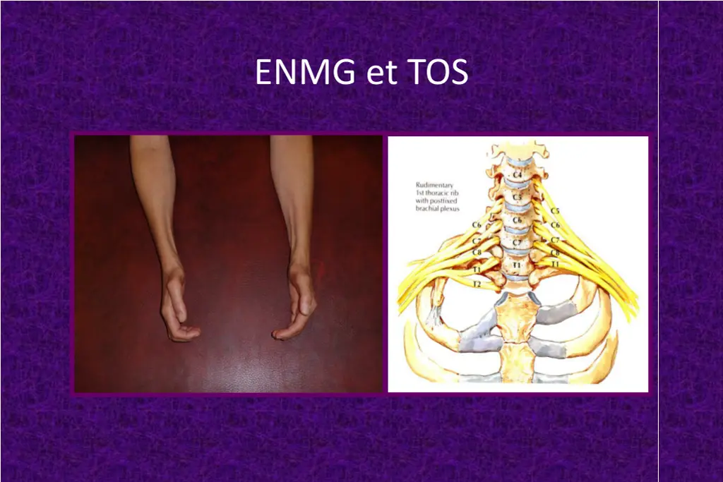
ENMG et TOS C:\Mes documents\DES2000\DES images\TOS neurologique.jpg
ENMG et TOS TOS SCC Oui Non Non Oui Non Oui/Non Oui Non C8 Oui Oui Oui Ulnaire coude Non Oui Oui Non Non Non Oui Non Non Oui Oui +++ Non Non Non Non Non Oui + Oui +++ Non Oui +++ Oui R d. Ampl PEM C.Abd.I R d. Ampl PEM Abd V R d. Ampl PEM 1er IO Aug. LDM m dian Aug. LDM ulnaire Alt ration m dian sensi. Alt ration ulnaire sensi. Alt ration BCI C.Abd.I neurog ne +++ Abd V et 1er IO neurog ne Oui/Non Non Non Non Oui Oui Oui Non
Neurographie sensitive Amp ( V) VC (m/s) M dian droit 48 53 M dian gauche 66 56 Ulnaire droit 21 57 Ulnaire gauche 17 60 BCI droit 0 BCI gauche 0 Neurographie motrice Amp (mV) F-M (ms) M dian droit 8,0 24,3 M dian gauche 3,4 29,3 Ulnaire droit 9,1 25,5 Ulnaire gauce 7,4 23,5
Ratio damplitude motrice ulnaire/m dian M dian moteur > Ulnaire moteur Ratio d amplitude motrice ulnaire/m dian (Lyu et al, 2011) [0.6 1.7] : sujets normaux > 1.7 : SLA (< 0.6 in 1/60), TOS < 0.6 : Maladie d Hirayama (34/46) 7,4 : 3,4 = 2,2
Early neurophysiological diagnosis of true neurogenic "thoracic outlet syndrome" (TOS) M.T. Hua, A. Dubuisson, B. Zeevaert, F.C. Wang (Li ge, B) Table 2 Patients and methods BARCELONA 2004 UNLIKELY TOS 1 2 3 4 5 6 7 Age Sex 54 56 36 51 44 53 47 MACN ( V) 11.0 9.6 16.0 0.9 10.0 12.0 9.1 Radial/MACN 1.5 3.1 3.7 - 1.8 2.4 4.8 F Uln - F Med (ms) -0.4 2.0 0.3 0.8 2.0 -0.7 0.2 X-ray normal normal normal normal normal normal Twenty-seven patients, diagnosed as having a TOS, were studied. The diagnosis was previously made by clinicians unrelated to our electrophysiological laboratory. Each patient was called back. History taking, physical examination and neurography evaluation were systematically performed. All had a cervical spine radiograph and a vascular-doppler flow study, otherwise the request was made. No other entrapment neuropathy was diagnosed in these 27 patients. According to their history, clinical features and vascular-doppler (Table 1), 3 groups were established : UNLIKELY TOS (n = 7, 4 women and 3 men, mean age = 48.7), VASCULAR TOS (n = 10, 8 women and 2 men, mean age = 41.6) and NEUROGENIC TOS with or without a vascular component (n = 10, 10 women, mean age = 44.8). We studied the medial antebrachial cutaneous nerve amplitude (MACN SNAP amplitude), radial SNAP amplitude/MACN SNAP amplitude ratio and the difference between minimal F-M latenties of median and ulnar nerves (F ulnar - F median) in these 3 groups (in bilateral TOS, only the worst side was considered) and in a group of 52 healthy subjects. Table 1 M W W M W M W VASCULAR TOS 1 2 3 4 5 6 7 8 9 10 Age Sex 38 44 52 39 35 48 45 33 26 56 MACN ( V) 4.1 15.0 11.0 8.4 8.7 6.2 9.1 12.0 12.0 5 Radial/MACN 6.8 2.7 3.0 2.6 6.1 3.5 3.5 3.4 2.8 2.2 F Uln - F Med (ms) 0.0 0.0 0.5 1.4 0.0 -0.5 1.8 0.4 0.7 0.5 X-ray sp normal normal normal normal normal + normal + + W W W M W W W W M W UNLIKELY TOS VASCULAR TOS NEUROLOGIC TOS - + - - /+ - - - - - + paresthesia : medial side of the upper limb weakness : hand intrinsic muscles sensory loss : medial side of forearm and/or hand strength decrease : thenar > hypothenar VASCULAR-DOPPLER + + + + - /+ Results NEUROLOGIC TOS 1 2 3 4 5 6 7 8 9 10 Age Sex 64 47 26 63 53 27 18 55 53 42 MACN ( V) 0.0 19.0 7.7 8.1 10.0 9.3 10.0 0.8 4.5 15.0 Radial/MACN - 3.0 4.3 5.9 2.4 4.0 5.1 26.3 4.0 2.5 F Uln - F Med (ms) -5.5 -0.6 0.3 0.0 1.0 -1.2 -0.5 -6.6 -0.3 -0.2 X-ray + normal normal normal normal + normal + + normal W W W W W W W W W W Control subjects: mean MACN amplitude was 18 5 V, lower limit of normal (LN) was 7.7 V ; mean amplitude ratio was 2 1 , upper LN was 3.8 ; mean F ulnar F median was 1.1 0.8 ms, lower LN was -0.4 ms (Table 3). Pathologic features were distributed as follows : 1) C7 transverse process hypertrophy or cervical rib : 0/7 case in UNLIKELY TOS, 2/10 cases in VASCULAR TOS and 4/10 cases in NEUROGENIC TOS ; 2) MACN amplitude < 7.7 V : 1/7 case in UNLIKELY TOS, 2/10 cases in VASCULAR TOS and 3/10 cases in NEUROGENIC TOS ; 3) amplitude ratio > 3.8 : 1/7 case in UNLIKELY TOS, 2/10 cases in VASCULAR TOS and 7/10 cases in NEUROGENIC TOS ; 4) F ulnar F median < -0.4 ms : 1/7 case in UNLIKELY TOS, 1/10 cases in VASCULAR TOS and 5/10 cases in NEUROGENIC TOS (Tables 2 and 3). Table 3 X-ray + = C7 transverse process hypertrophy or cervical rib MACN : medial antebrachial cutaneous nerve Uln : ulnar ; Med : median ; M : man ; W : woman X-ray + MACN ( V) 18 5 >/= 7.7 9.8 4.6 1 9.6 3.1 2 8.4 5.8 3 Radial/MACN amplitude ratio 2 1 </= 3.8 2.5 1.6 1 3.7 1.5 2 6.4 7.5 7 F Uln - F Med (ms) 1.1 0.8 >/= -0.4 0.6 1.1 1 0.5 0.7 1 -1.4 2.6 5 Healthy subjects (n = 52, M/W = 30/22) (mean age = 29.0) UNLIKELY TOS (n = 7, M/W = 5/3) (mean age = 48.6) VASCULAR TOS (n = 10, M/W = 8/2) (mean age = 41.6) NEUROLOGIC TOS (n = 10, M/W = 10/0) (mean age = 44.8) Mean SD Limit of normal Mean SD Number of pathologic cases Mean SD Number of pathologic cases Mean SD Number of pathologic cases Conclusion 0/6 Our results suggest that the amplitude ratio (radial SNAP amplitude/MACN SNAP amplitude) is more sensitive than the MACN amplitude for the diagnosis of TOS. The difference between F- wave latenties of median and ulnar nerves (F median 0.4 ms longer strengthen the presumption of TOS, as far as there is no associated carpal tunnel syndrome. 3/10 than F ulnar) can 4/10
Nouvelle neurographie sensitive Branche terminale sensitive du nerf radial : - 8 cm entre stimulation et d tection BCI/nerf cutan antebrachial m dial : - 8 cm entre stimulation et d tection - 4 cm de part et d autre du pli du coude
Nouvelle neurographie sensitive N=50 Moyenn e M dian e ET Min Max LN Age 44 14,3 10 93 Taille 168 9,4 145 192 VC radial 62 63 6,1 46,8 72,7 52,4 Amp radial 48 50 14,5 17 84 24,5 VC BCI 66 66 5,8 55,6 80 56,6 Amp BCI Radial/BCI 20 2,5 20 2,6 6,4 0,8 10 1 42,7 4,4 9,5 3,8