Neuroprotective Effects of Exercise on Parkinson's Model
In Parkinson's disease, the basal ganglia play a crucial role. Understanding the organization and connections within the basal ganglia sheds light on the disease pathology. This study explores the neuroprotective impact of exercise in a rat model treated with rotenone, a toxin that induces Parkinsonism. The intricate neural circuitry of the basal ganglia, involving various nuclei and neurotransmitters, influences motor functions. The interconnections within the basal ganglia, such as the nigrostriatal projections and inhibitory pathways, are crucial in motor control. Investigating these mechanisms provides insight into potential therapeutic strategies for Parkinson's disease.

Download Presentation

Please find below an Image/Link to download the presentation.
The content on the website is provided AS IS for your information and personal use only. It may not be sold, licensed, or shared on other websites without obtaining consent from the author.If you encounter any issues during the download, it is possible that the publisher has removed the file from their server.
You are allowed to download the files provided on this website for personal or commercial use, subject to the condition that they are used lawfully. All files are the property of their respective owners.
The content on the website is provided AS IS for your information and personal use only. It may not be sold, licensed, or shared on other websites without obtaining consent from the author.
E N D
Presentation Transcript
THE NEUROPROTECTIVE EFFECTS OF EXERCISE ON ROTENONE-TREATED RAT MODEL OF PARKINSON'S DISEASE
Organization of basal ganglia The term basal ganglia applied to 5 interactive strucures on each side of the brain. Caudate nucleus. Putamen. Globus pallidus. Subthalamic nucleus. Substantia nigra.
Organization of basal ganglia Caudate nucleus + Putamen = stiatum. Putamen + Globus pallidus = lenticular nucleus. Globus pallidus is devided into internal &external segments(Gpi &Gpe) GABAergic neurons. Substantia nigra Pars Compacta (dopaminergic) &Pars Reticularis (GABAergic)
Organization of basal ganglia At least there are 4 types of striatal neurons. 95% of striatal neurons medium spiny neurons (GABA). Aspiny neurons: - Large : Acetylcholine. - Medium: Somatostatin. - small:GABA.
The Basal Ganglia connections (Input) Input : 2 major inputs both are exitatory (glutamate) & both terminate on the stiatum, which are: -corticostriate pathway. - Thalamostriatal pathway: from intralaminar nuclei of the thalamus.
The Basal Ganglia connections (Output) Output : (GABA)& both project to the thalamus, - SNPR - Gpi From the thalamus projections to prefrontal & premotor cortex. This completes a full cortical-basal ganglia-thalamic- cortical loop(putamen circuit and caudate circuit). 2 major output of both are inhibitory exitatory (may be glutamate)
The Basal Ganglia connections (Interconnections) Nigrostriatal projection (dopaminergic) from SNPC to the striatum GABAergic projection from striatum to SNPR. Inhibitory projections from striatum to Gpi &GPe. Gpe send inhibitory projection to subthalamic nuclei in turn send exitatory projections (Glutamate) to Gpe &Gpi. 1. 2. 3. 4.
Brain stem connections of basal ganglia Fibers from Gpi PPN (pedunculopontine nuclei, part of reticular formation) brain stem and spinal cord. Vestibular Nucleus, Red Nucleus & Inferior Olivary Nucleus receive projections fom GPi then to the spinal cord through extrapyramidal tracts.
Basal Ganglia Pathways a) Direct pathway Activated by binding of dopamine to D1 receptors in the striatum. Direct inhibition of basal ganglia output to motor thalamus (inibitory output) Disinhibition of motor thalamus promote movement. Lesion difficulty intiating movement ( PD).
Basal Ganglia pathways b) Indirect pathway Inhibited by bining of dopamine to D2 receptors in the striatum. Indiect stimulation of basal ganglia inhibitory output to motor thalamus subthalamic nucleus. Inhibition of motor thalamus inhibition of movement. Lesion spontaneous movement (Huntington disease).
Basal Ganglia function 1- Planning and programming of movement. 2- Control of automatic associated movements. 3- Caudate nucleus play role in some cognitive processes. Through its inter-connection with frontal portion of the neocortex. Lesion in caudate N. disrupt test involving object reversal & delayed alternation Lesions of left(not right) head of caudate N. dysarthic form of aphasia resembles wernicke aphasia. 4- Control of muscle tone . Lentiform N. decrease MT inhibit vestibular N.& stimulate the inhibitory reticular formation. Caudate N. facilitate MT stimulate facilitatory reticular formation ,vestibular &olivary N. - - - -
Basal Ganglia Diseases 3 distnict biochemical pathways operate in balance , lesion in one or more abnormality 1- nigrostriatal dopaminergic system. 2- intrastriatal cholinergic system. 3- GABAergic system from the striatum ti SN & GP. 2 types of disorders , hyperkinetic (chorea, asthetosis & ballism) or hypokinetic (akinesia & bradykinesia..
Basal Ganglia diseases Chorea. Asthetosis. Hemiballismus. PD. Wilson disease. Kernicterus. Tardive dyskinesia. 1. 2. 3. 4. 5. 6. 7.
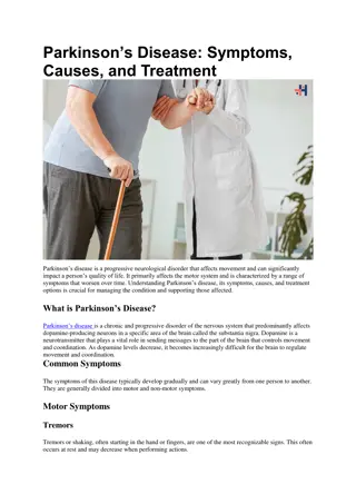
Parkinson disease(PD) PD is a degenerative disease involving loss of dopaminergic neurons in the substantia nigra, which cause relative deficiency of dopamine and excess of acetyl ch. in the striatum. It was originally described in 1817 by James Parkinson and is named for him. Parkinson disease is the first disease identified as being due to a deficiency in a specific neurotransmitter .In the 1960s, Parkinson disease was shown to result from the degeneration of dopaminergic neurons in the substantia nigra pars compacta. The fibers to the putamen (part of the striatum) are most severely affected.
There are 710 million people worldwide in whom Parkinson disease has been diagnosed. The disease is 1.5 times more prevalent in men than women. Parkinsonism occurs in sporadic idiopathic form in many middle-aged and elderly individuals and is one of the most common neurodegenerative diseases. It is estimated to occur in 1 2% of individuals over age 65. Dopaminergic neurons and dopamine receptors are steadily lost with age in the basal ganglia in healthy individuals, and an acceleration of these losses apparently precipitates parkinsonism. Symptoms appear when 60 80% of the nigrostriatal dopaminergic neurons degenerate.
Etiology of PD the loss of dopamine (DA) neurons in the substantia nigra (SN) is responsible for most of the characteristic motor dysfunctions, and there is reason to believe that the loss of DA contributes to other aspects of the disorder, as well. Environmental toxins are major risk factors. Mitochondrial dysfunction is central to the pathophysiology of PD. decreased mitochondrial complex I activity and increased activity of the ?-ketoglutarate dehydrogenase complex in patients with PD. Oxidative stress which can result from mitochondrial dysfunction, reduced antioxidant capacity, and reactive oxygen species that occur due to DA metabolism also plays a significant role in the etiology of the disease. There is a significant inflammatory component of the disease. 1. 2. 3. 4. 5.
Features of PD PD has both hypokinetic and hyperkinetic features. The hypokinetic features of Parkinson disease are akinesia and bradykinesia. the hyperkinetic features are cogwheel rigidity and tremor at rest.
Manifestation of PD Muscle rigidity 1. The rigidity is different from spasticity because motor neuron discharge increases to both the agonist and antagonist muscles. Passive motion of an extremity meets with a plastic, dead-feeling resistance that has been likened to bending a lead pipe and is therefore called lead pipe rigidity. Sometimes a series of catches takes place during passive motion (cogwheel rigidity), but the sudden loss of resistance seen in a spastic extremity is absent.
2- Akinesia or dyskinesia(lack of movement)and bradykinesia Absence of motor activity and the difficulty in initiating voluntary movements. There is a decrease in the normal unconscious movements such as swinging of the arms during walking. lack of facial expressions (musk face). Slow monotonus and low volume speech. Shuffling gait.
3- Static Tremors The tremor, which is present at rest and disappears with activity and durind sleep, is due to regular, alternating 8-Hz contractions of antagonistic muscles. Marked in the upper limb, in the hand (pill-rolling movement).
Pathogenesis of PD In healthy individuals, basal ganglia output is inhibitory via GABAergic nerve fibers. The dopaminergic neurons that project from the substantia nigra to the putamen normally have two effects. They stimulate the D1 dopamine receptors, which inhibit GPi via direct GABAergic receptors; and they inhibit D2 receptors, which also inhibit the GPi. the inhibition reduces the excitatory discharge from the subthalamic nucleus to the GPi. This balance between inhibition and excitation somehow maintains normal motor function.
In PD, the dopaminergic input to the putamen is lost. This results in decreased inhibition and increased excitation from the subthalamic nucleus to the GPi. The overall increase in inhibitory output to the thalamus and brainstem disorganizes movement. The genes for at least five proteins can be mutated. These proteins appear to be involved in ubiquitination. Two of the proteins, -synuclein and barkin, interact and are found in Lewy bodies. The Lewy bodies are inclusion bodies in neurons that occur in all forms of Parkinson disease.
An important consideration in Parkinson disease is the balance between the excitatory discharge of cholinergic interneurons and the inhibitory dopaminergic input in the striatum. Some improvement is produced by decreasing the cholinergic influence with anticholinergic drugs. More dramatic improvement is produced by administration of L-dopa (levodopa).
Treatment of PD There is no cure for Parkinson disease, and drug therapies are designed to treat the symptoms. Improvement is produced by administration of L-dopa (levodopa). Unlike dopamine, this dopamine precursor crosses the blood-brain barrier and helps repair the dopamine deficiency. However, the degeneration of these neurons continues and in 5 7 years the beneficial effects of L-dopa disappear. Sinemet, a combination of levodopa (L-dopa) and carbidopa, is the most commonly used drug for the treatment of Parkinson disease. The addition of carbidopa to L-dopa increases its effectiveness and prevents the conversion of L-dopa to dopamine in the periphery and thus reduces some of the adverse side effects of L-dopa (nausea, vomiting, and cardiac rhythm disturbances).
Dopamine agonists, including apomorphine, bromocriptine, pramipexole, and ropinirole, have also proven effective in some patients with Parkinson disease. As combination with levodopa, cathechol-O- methyltransferase (COMT) inhibitors (eg, entacapone) are another class of drugs used to treat this disease. They act by blocking the breakdown of L-dopa, allowing more of it to reach the brain to increase the level of dopamine. MAO-B inhibitors (eg, selegiline) also prevent the breakdown of dopamine. They can be given soon after diagnosis and delay the need for levodopa.
The US Food and Drug Administration has approved the use of deep brain stimulation (DBS) as a method for treating Parkinson disease. DBS reduces the amount of L-dopa patients need and thus reduce its adverse side effects (eg, involuntary movements called dyskinesia). DBS has been associated with reducing tremors, slowness of movements, and gait problems in some patients.
Surgical treatment of PD Surgical treatments in general are reserved for those who have exhausted drug therapies or who have not responded favorably to them. Lesions in GPi (pallidotomy) or in the subthalamic nucleus (thalamotomy) have been performed to help restore the output balance of the basal ganglia toward normal . Another surgical approach is to implant dopamine-secreting tissue in or near the basal ganglia. Transplants of the patient s own adrenal medullary tissue or carotid body works for a while, apparently by functioning as a sort of dopamine minipump, but long-term results have been disappointing. Results with transplantation of fetal striatal tissue have been better, and there is evidence that the transplanted cells not only survive but make appropriate connections in the host s basal ganglia. However, some patients with transplants develop dyskinesias due to excessive levels of dopamine.
Animal models of PD There are three current techniques for producing in vivo models of Parkinson s disease: 1-Unilateral lesioning with 6-hydroxydopamine (rats) 2-Systemic injection of 1-methyl 4- phenyltetrahydropyridine (MPTP)(mice and monkeys). 3-Systemic injection of rotenone (rats).
6-OHDA RAT MODEL This is the traditional model for testing Parkinson s therapies, especially those intended to increase dopamine levels in the striatum. The principal advantage of this model is that it is very sensitive to dopamine agonists. The toxin 6-hydroxydopamine (6-OHDA) is injected on one side of the rat, while the opposite side serves as an intra-animal control. This injection produces DA neuron loss on the injected side while sparing the contralateral DA neurons.
MPTP MOUSE MODEL The MPTP mouse model of Parkinson s disease is thought to mimic more closely the behavioral pathology of Parkinson s disease, compared to the 6-OHDA rat model, and is currently the model of choice. Mice exhibit Parkinson s-like symptoms following systemic injection of the pyridine toxin MPTP, which produces a loss of striatal dopamine (DA) nerve terminal markers and, at higher doses, death of DA neurons in the substantia nigra. Since the process of terminal loss and degeneration takes 6- 9 days following MPTP injection, drugs can be administered both before the MPTP is given (to assay neuroprotection), and/or while the toxin is active in the first week post- injection (to assay preservation of dopamine function).
THE ROTENONE MODEL The most recent of the Parkinson s models uses systemic injection of rotenone, a member of a class of compounds found in pesticides, and in natural products, that disrupts mitochondrial activity and results in pathology that is strikingly similar to that seen in Parkinson s disease (e.g., accumulation and aggregation of -synuclein and ubiquitin, progressive oxidative damage, and caspase dependent death).
Only recently have reliable behavior tests become available with this model. This reliability comes from using two distinct protocols, each with different optimal doses of the toxin. In the first protocol, a lower dose is used that minimizes the loss of animals before the 3-week endpoint of a typical study, yet still produces a decrease in TH in the striatum. The extent of reduced TH activity is variable across rats and there is no correlation between the extent of TH loss and the behavioral deficits. Thus, this model is appropriate for testing compounds that reduce toxicity and the resultant behavioral deficits, but their effect on dopamine activity is secondary.
In the second protocol a higher dose of rotenone is used that produces pronounced neuropathology. In this case the endpoint measure is acute toxic behavior that requires perfusion of the animal. These animals have a reliable loss of TH in the striatum and loss of DA neurons in the nigra, both of which can be measured. A neuroprotective compound tested in this version of the model would decrease the number of rats displaying toxic behavior at a set post-lesion time point, or prolong the average time before toxic behavior. A positive control in this protocol is Coenzyme Q, which is neuroprotective.
MPTP PRIMATE MODEL Similar to the MPTP mouse model, this model is most often used as a final study of a test compound s efficacy before testing in humans. The non-human primate species used are either cynomolgus, rhesus, or African green monkeys. Following systemic MPTP administration, the animal s motor and cognitive functions are impaired, as in humans. In particular the animals display muscle rigidity, akinesia, and tremors, along with impairment on a common memory task, delayed match to sample (DMTS).
IN VITRO MODELS Rotenone A botanical compound obtained from the roots of Derris sp., rotenone is an active ingredient in many pesticides. Rotenone blocks complex I of the mitochondrial respiratory chain and there by induces nigrostriatal degeneration in rodents, thus reproducing the hallmark pathology of Parkinson s disease. MPP+ The active metabolite of MPTP, MPP+ is a piperidine derivative which induces a Parkinsonian syndrome in mice and primates. MPP+ selectively enters dopamine neurons via the dopamine transporter and also blocks complex I of the mitochondrial respiratory chain. Epoxymicin Epoxymicin, a natural product isolated from Actinomyces sp., is a cell permeable, potent, selective and irreversible proteasome inhibitor. Impairment of the ubiquitin-proteasome system replicates features of Parkinson s disease.
Exercise and PD For many years, there has been the notion that an altered environment can have an impact on neuronal structure and function. One of the earliest mentions of this idea was by the Italian neuroanatomist Michele Vicenzo Malacarne, who reported in 1793 that dogs and birds that had undergone training had larger and more complex cerebellar structures than unattended littermates. In the early 1800s, Johann Spurzheim suggested that the brain was capable of increasing in size due to exercise and proposed that this idea be tested by a rigorous application of the scientific method. The idea that the size of the brain could be altered in response to changes in the environment, however, stayed within the purview of phrenologists for over 100 years.
In 1947, Donald Hebb described superior maze performance of rats reared as domestic pets compared with their relatively impoverished laboratory-reared counterparts. Then, in the 1960s,Krech, Bennett, and Rosenzweig, The team housed groups of rats in a large cage that provided many opportunities to explore the space and the objects it contained, some of which were frequently changed. They found that this led to increased social interaction, physical exercise, and opportunities to learn that were associated with changes in the structure, neurochemistry, and function of the rodent brain.
Physical exercise is currently applied as a behavioral intervention to improve motor symptoms in patients with PD (Lee et al., 2015). Exercise can improve and alleviate memory loss in elderly(Karmer et al., 2006),and decrease the risk of developing PD (Cotman and Berchtold,2002).the neuroprotective potentials of exercise is great, but the underlying mechanisms remain a debatable issue.
Putative Mechanisms Underlying the Relationship of Physical Activity and Cognitive Function Four main hypotheses have been proposed to explain these mechanisms: The cardiovascular. Immunologic. Neuroendocrine. Neurotrophic signaling hypothesis. 1. 2. 3. 4.
Cardiovascular Effects Adequate levels of physical activity are vital for improved small vessel condition (Schmidt et al., 2013), increased cerebral blood flow, and nutrient delivery (Swain et al., 2003). Increased levels of physical activity were inversely correlated with reductions in gray and white matter loss, particularly in the prefrontal, superior parietal, and temporal cortices (Colcombe et al., 2003).
Rodent studies have demonstrated that running exercise lead to improvements in vascular perfusion and angiogenesis in the motor cortex, alterations that were restricted to the motor regions of the cerebral cortex. Other studies have suggested that motor skill training increases synaptogenesis in corresponding regions (Kleim et al., 1996)
aged individuals accrue cognitive deficits over time. physical activity is an important modulator of cognition across the lifespan, but that the contribution of cardiovascular fitness for sustaining healthy cognitive function increases with age by impacting central and peripheral cellular processes.
Immunological Effects Physical activity results in increased levels of pro- inflammatory, anti-inflammatory cytokines, cytokine inhibitors and chemokines depending upon the intensity and duration of such exercise, with overall enhancement of immune function and anti-inflammatory processes. Regular physical activity decrease viral and bacterial infections (DiPenta et al., 2004; Kohut and Senchina, 2004), a lower incidence of systemic lowgrade inflammation (Stewart, 2002; Colbert et al., 2004), and a lower incidence of neurodegeneration and cognitive decline (Cotman et al., 2007).
Kohut et al. (2006) studied older adults participating in aerobic or flexibility exercise for 10 months and found a significant reduction in plasma levels of interleukin 6 (IL-6), interleukin 8 (IL-8), C-reactive protein (CRP), and tumor necrosis factor (TNF), linking physical activity to antiinflammatory processes in the central nervous system (CNS).
TROPHIC FACTOR SIGNALING Neurotrophins Improved trophic factor signaling has been considered as the most popular hypothesis to explain the positive effects of physical activity on cognition, with attention centering on the neurotrophins (NTs). NTs are comprised of a closely related family of polypeptides that regulate a variety of neuronal functions including proliferation, survival, migration, and differentiation (Salehi et al., 2003, 2004). The mammalian NTs include nerve growth factor (NGF), brain derived neurotrophic factor( BDNF), neurotrophin-3 (NT-3) and, neurotrophin- 4/5 (NT4/5). These factors are synthesized by target neurons, initially in the form of pre-pro-protein.


")


 studied older adults")