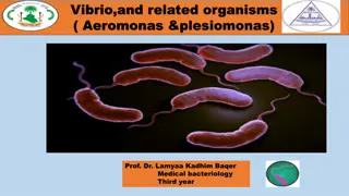
Non-spore forming Gram-positive bacilli Corynebacterium
The presentation delves into the world of die materials in the field of conservative dentistry and endodontics, covering ideal requisites, classification, systems, and more. Detailed learning objectives are set to enhance understanding.

Uploaded on Feb 24, 2025 | 1 Views
Download Presentation

Please find below an Image/Link to download the presentation.
The content on the website is provided AS IS for your information and personal use only. It may not be sold, licensed, or shared on other websites without obtaining consent from the author.If you encounter any issues during the download, it is possible that the publisher has removed the file from their server.
You are allowed to download the files provided on this website for personal or commercial use, subject to the condition that they are used lawfully. All files are the property of their respective owners.
The content on the website is provided AS IS for your information and personal use only. It may not be sold, licensed, or shared on other websites without obtaining consent from the author.
E N D
Presentation Transcript
Non-spore forming Gram-positive bacilli Corynebacterium Members of the genus Corynebacterium are Actinomycetes that are related to he genera Mycobacteriun, Nocardia and Rhodococcus. Corynebacterium pleomorphic rods often with coccoid or club-shaped appearance. C. diphtheria is the agent of human diphtheria. They are aerobic or facultatively anaerobic, catalase +, non-spore forming & non- acid fast, with little tendency to branch. C. diphtheria grow aerobically on most ordinary media. On blood agar the colonies are small, granular and gray & may have small zone of hemolysis. On agar containing potassium tellurite, the colonies are brown to black with surrounded by halo. In stained smear from culture medium the bacilli arranged in chines letter appearance. On selective Tinsdale media, black colonies surrounded by halo (Diagnostic). are aerobic G+
Infections caused by Corynebacterium C. diphtheriae & C. ulcerans are prominent human pathogen. C. diphtheriae & C. ulcerans can cause bovine mastitis and dermatitis. animal pathogens are C. pseudotuberculosis , a cause of lymphadenitis & lymphangitis in small ruminants and horses. C. renale( C. cystitidis, C.pilosum) are opportunistic agents of UTIs in cattle & occasionally C.pseudotuberculosis & C. ulcerns can produce diphtheria toxin. The notable some other species.
Diseases, Epidemiology & Pathogenesis C. renale is a normal flora of the lower urogenital tract. C.pilosum is also normal flora, & is less common cause of cystitis & pyelonephritis. C. cystitidis is usually associated with chronic pyelonephritis, but can cause more severe cystitis than the other members. The members of the C.renale group are opportunistic pathogens of UT of cattle and other domestic animals. The major risk factors; shortness of the female urethra, pregnancy and parturition, anatomic anomalies, physical damage & obstruction of UT. The disease occur most frequently in mature cows, & to 1/3 of cases are fatal.
Diseases, Epidemiology & Pathogenesis Colonization is pilus mediated & the rapidly urease + nature of the bacteria lead to production of ammonia with resulting mucous inflammation. Cows with alkaline urine are at greater risk for developing pyelonephritis. The clinical presentation of acute pyelonephritis includes fever, anorexia, polyuria, hematuria, pyuria & abdominal posture (arched back). Chronic pyelonephritis associated with weight loss, anorexia & decreased milk production. The first step in the pathogenesis of pyelonephritis is attachment of the bacteria to the urethral epithelium. Bacteria grow readily causing cystitis & ascending to the kidney. The virulence factor of C.renale is Renalin (an extracellular protein) that play a role in lysis of cell membranes. This protein is not produced by C.pilosum or C.cystitidis. Pilli are produced by the three bacteria. Piliated bacteria are more resistant to phagocytosis.
C.Pseudotuberculosis is a facultative intracellular C.pseudotuberculosis bacteria that is the cause of caseous lymphadenitis (CLA) in goats & sheep, ulcerative lymphangitis & ventral abscess in horses, and abscess and mastitis in cattle. CLA is economically significant worldwide (usually due to death), characterized by chronic abscessation of peripheral LNs. Prevalence rate of 30%-50% may occur in herds or flocks, & higher rate with older age. Transmission occurs primarily though direct contact (Aerosol). Bacteria may enter through shearing wounds & abrasions. Phagocytosed bacteria are transported to regional LN leading to abscess formation. Dissemination from primary to secondary sites may occur.
C.ulcerns is best known as a cause of pharyngitis in human. In animals causes of bovine mastitis. C.ulcerns are able to produce diphtheria toxin. Infected cows may shed the bacteria with the milk for months or years & sporadic human cases are associated with consumption of raw milk (50% of C.ulcerns isolates from the milk produce toxin). C. Ulcerns C.bovis diphtheria C.bovis udder where it attached to epithelium of teat duct. 20% of cattle carry C.bovis. Growth of bacteria is enhanced in the milk during lactation. Inflammatory changes may occur in teat canal & mammary paranchyma. is a commensal of bovine
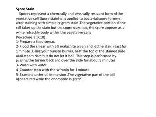
* Specimens: throat swab, wound swab, pus from abscess, milk, urine, genital secretion. *Direct stain: Grams stain or Albert stain (deep blue metachromatic granules & faint green shaft). Culture on ordinary & selective media: Blood agar, potassium telurite blood agar & Tensdale medium (selective media). Colony morphology & identification: Albert stain Toxigenicity test: Detection of diphtheria toxin Laboratory diagnosis