OPERATING MICROSCOPE - PHYSICS, OPTICS AND USES IN NEUROSURGERY
The operating microscope plays a crucial role in neurosurgery by providing magnified views of the surgical site. It utilizes basic optics to achieve magnification and stereoscopic vision, enhancing the precision of procedures. The history of its development, including key milestones in the use of magnification loupes and surgical binocular microscopes, reflects significant advancements in microsurgery. Optical principles such as magnification and field of view are fundamental to the functionality of the operating microscope, enabling surgeons to perform intricate procedures with enhanced visual clarity.

Download Presentation

Please find below an Image/Link to download the presentation.
The content on the website is provided AS IS for your information and personal use only. It may not be sold, licensed, or shared on other websites without obtaining consent from the author.If you encounter any issues during the download, it is possible that the publisher has removed the file from their server.
You are allowed to download the files provided on this website for personal or commercial use, subject to the condition that they are used lawfully. All files are the property of their respective owners.
The content on the website is provided AS IS for your information and personal use only. It may not be sold, licensed, or shared on other websites without obtaining consent from the author.
E N D
Presentation Transcript
OPERATING MICROSCOPE - PHYSICS, OPTICS AND USES IN NEUROSURGERY
Basic Optics Basic function of a microscope is to provide a magnified view of the object being studied Magnification is essentially an increase in viewing angle or the angle subtended by the object at the eye. Two essentials of the operating microscope Magnification Stereoscopic vision
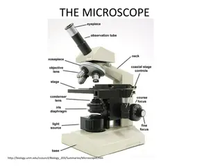
Simple microscope Consists of an illumination source and two lens system Objective lens Eyepiece The objective lens focuses the light rays from the object under study to form a real inverted image The eyepiece forms a virtual magnified image at a distance which is seen by the observer. The image undergoes a two step magnification.
Magnifying power of a microscope is calculated by multiplying the individual magnification produced by the objective lens and eyepiece individually. Simple lens systems suffer from several defects such as chromatic aberration, Spherical aberration, diffraction Compound lens systems diminish these deficiencies
The operating microscope History of its development magnification loupes have been used in surgery from mid part of 19thcentury. Surgical binocular microscope first used by Carl Nylen in 1929 for middle ear surgery. Popularised for otological surgery by William House In 1957, Theodore Kurze became the first neurosurgeon to use the microscope in removal of a neurilemmoma of the seventh nerve.
History (contd..) In 1958, R.M.P. Donaghy established the first microneurosurgical training laboratory where several neurosurgeons like M Gazi Yasargil also trained Yasargil made several revolutionary improvements in the design of the operating microscope and is regarded as the Father Of Microneurosurgery for his contributions.
The operating microscope Optical principles Magnification : dependent on the magnification of the objective and eyepiece a zoom system of lenses is interposed between these two principal lenses allowing continuous change in magnification. The field of view changes with the magnification according to the formula- Diameter of field = 200/total magnification
Depth of field is also an important parameter which is a measure of field of vision in a stereoscopic system. The depth of field increases with the square of the focal length of the objective lens decreases linearly with the magnification of the microscope.
Components Of An Operating Microscope Main Objective Lens variable focal length ranging from 200-500 mm depending upon the depth of operative field allowing the microscope to be adjusted at different distances from the op cavity. Greater focal length required for operating in depth. Magnification Changer It is a lens system placed between the objective and the binocular system comprising microprocessor controlled lenses which allow continuous adjustment of magnification Together with the objective lenses they form the double barrel system.
Illumination Earlier microscopes used integrated light sources such as tungsten or halogen bulbs which generated a lot of heat. Prolonged surgery cumbersome. Development of fibreoptics enabled the use of a remote illumination source . Automatic adjustment of light collimation in modern microscopes allows appropriate illumination as the magnification is varied.
Auxiliary illumination In some advanced models auxiliary illumination is being used to decrease shadowing when changing the viewing angle.
Stereoscopic perspective Each of the binocular eyepieces project a slightly different image of the field which is fused to form the resultant 3D image. The binocular system ensures that the two images are always separated by the interpupillary distance of the observer irrespective of the depth of the field.
Operative Microscope-based Neuronavigational Systems Neuronavigation provides a precise surgical guidance by referencing the coordinate system of the brain with a parallel coordinate system of the three-dimensional data of the patient . Picture in picture facility : the simultaneous display of the image data into the eyepiece of the microscope from either the neuronavigational system or during the use of an intraoperative endoscope is possible.
Microscope mounts Essentially two types Floor mounted transportable, occupies floor space Ceiling mounted More expensive, saves floor space. Unnecessary movements microscope counterbalances and electromagnetic locks which secure the microscope in the desirable position. while adjusting a system the of are minimised by
Point lock system and focus lock system available in advanced models The use of the point lock mechanism allows the surgeon to position the microscope without any chance of losing the observation point or the focus of that point. focus lock allows the surgeon to position the microscope in an x-y plane without affecting the z axis
Extent and scope of application of microscope in neurosurgery. Role of microscope in improving surgical outcomes first demonstrated in Acoustic Neuromas. Now routinely used in almost all intradural operative procedures whether in the brain or spine. Its use has resulted postoperative neural and vascular damage, better hemostasis, more accurate nerve and vessel repairs, and surgical treatment of some previously inoperable lesions. in smaller wounds, less
It has improved operative results by permitting neural and vascular structures to be delineated with greater visual accuracy deep areas to be reached with less brain retraction and smaller cortical incisions bleeding points to be coagulated with less damage to adjacent neural structures, nerves distorted by tumor to be preserved with greater frequency and enabling anastomosis and suturing of small vessels and nerves not previously possible to be performed.
Emerging technologies Intraoperative flourescence It is an upcoming technique available in several advanced micrscopes. applicable in aneurysm and tumour surgery where it allows the visualisation of sub millimeter vessels by the use of Indo-cyanin green dye used as fluorescing agent


")