Optical Clearing Using Mannitol: A Confocal Raman Micro-spectroscopy Study on Porcine Dura Mater
Explore the optical clearing of ex-vivo porcine dura mater using mannitol through confocal Raman micro-spectroscopy. This study investigates the mechanisms and benefits of optical clearing in tissue diagnosis and treatment, highlighting the reduction of light scattering and enhancement of optical diagnostic methods. Learn about the main mechanisms proposed for tissue immersion optical clearing and the noninvasive optical techniques implemented for deep biological investigation. Discover the hypothesized mechanisms of optical clearing and the materials and methods used in confocal Raman microspectroscopy. Explore the potential applications of optical clearing in improving contrast and spatial resolution in optical diagnostic methods.
Download Presentation

Please find below an Image/Link to download the presentation.
The content on the website is provided AS IS for your information and personal use only. It may not be sold, licensed, or shared on other websites without obtaining consent from the author. If you encounter any issues during the download, it is possible that the publisher has removed the file from their server.
You are allowed to download the files provided on this website for personal or commercial use, subject to the condition that they are used lawfully. All files are the property of their respective owners.
The content on the website is provided AS IS for your information and personal use only. It may not be sold, licensed, or shared on other websites without obtaining consent from the author.
E N D
Presentation Transcript
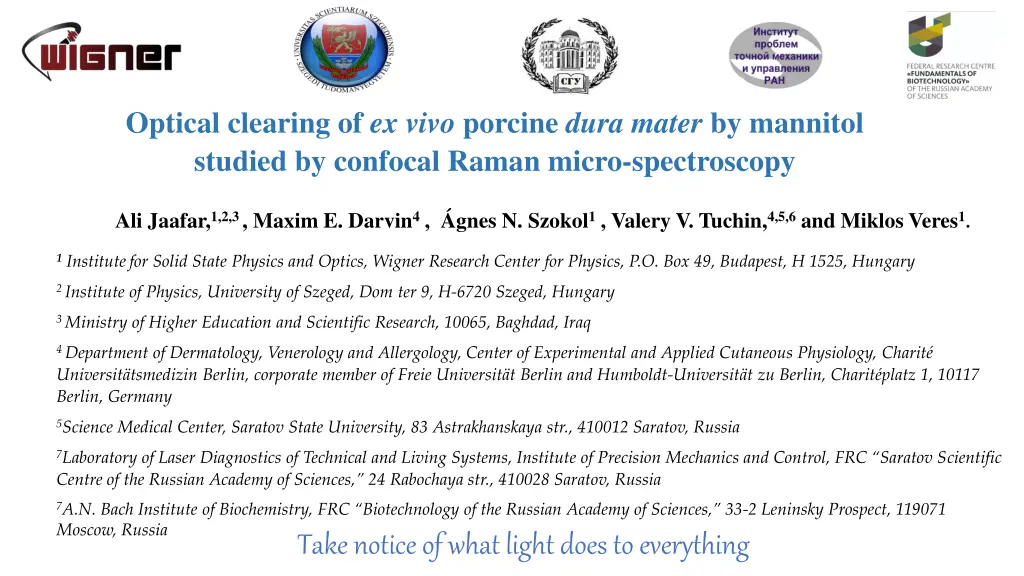
Optical clearing of ex vivo porcine dura mater by mannitol studied by confocal Raman micro-spectroscopy Ali Jaafar,1,2,3 , Maxim E. Darvin4 , gnes N. Szokol1 , Valery V. Tuchin,4,5,6 and Miklos Veres1. 1Institutefor Solid State Physics and Optics, Wigner Research Center for Physics, P.O. Box 49, Budapest, H 1525, Hungary 2 Institute of Physics, University of Szeged, Dom ter 9, H-6720 Szeged, Hungary 3 Ministry of Higher Education and Scientific Research, 10065, Baghdad, Iraq 4 Department of Dermatology, Venerology and Allergology, Center of Experimental and Applied Cutaneous Physiology, Charit Universit tsmedizin Berlin, corporate member of Freie Universit t Berlin and Humboldt-Universit t zu Berlin, Charit platz 1, 10117 Berlin, Germany 5Science Medical Center, Saratov State University, 83 Astrakhanskaya str., 410012 Saratov, Russia 7Laboratory of Laser Diagnostics of Technical and Living Systems, Institute of Precision Mechanics and Control, FRC Saratov Scientific Centre of the Russian Academy of Sciences, 24 Rabochaya str., 410028 Saratov, Russia 7 .N. Bach Institute of Biochemistry, FRC Biotechnology of the Russian Academy of Sciences, 33-2 Leninsky Prospect, 119071 Moscow, Russia Take notice of what light does to everything
Optical clearing Main mechanisms of tissue optical immersion clearing Materials and methods: Confocal Raman microscopy Dura mater sample preparation Optical Clearing Agents Results: optical clearing using confocal Raman micro- spectroscopy Conclusions
Optical clearing Noninvasive optical techniques implementation in tissue diagnosis and treatment, such as confocal Raman Raman micro-spectroscopy, coherent anti- Stokes Raman scattering (CARS) spectroscopy and tomography, multiphoton tomography (MPT), SHG- microscopy and OCT. Main limitation of the deep biological investigation that all tissue layers are characterized by high light scattering and absorption Since it was reported in the nineties, the optical clearing (OC) technique was developed to allow effectively reduce light scattering in tissue to reach the maximum probing depth, improve contrast and spatial resolution of optical diagnostic methods. Schematically shows the distribution of mannitol molecules when a dura mater sample is immersed in glycerol
SPIE Press 2005 Three hypothesized mechanisms of OC were suggested: Main mechanisms of tissue immersion optical clearing (OC) Matching of refractive indices between tissue components and interstitial fluid Tissue dehydration Reversible dissociation of collagen fibers These and possibly other not known OC mechanisms usually work not independently but simultaneously with different relative contributions dependent on tissue and OCA and delivery method. Springer 2019 CRC Press 2022
Materials and methods: Confocal Raman microspectroscopy The excitation source: laser: 633 nm, power: 8.3 mW, exposure time 5 sec, Grating: 1200, spectral range: 400 to 1800 cm 1 , 50 objective and was used to focus the laser beam and to collect Raman signals Measurement protocol of sample OC: Measurements were performed on fresh ex vivo porcine Dura mater (DM). Treatment time was for 1, 2, 3, 5 and 10 min immersion in mannitol bath of petri dish with concentration 0.16 g/ml, the refractive index was 1.354. Series of Raman spectra obtained from the DM at different depths ranging (from 0 to 125 m) with 25 m step size. Before measurements, mannitol was removed from samples surface using a paper towel Sample size 19 mm, thickness 400 m approx., 5 samples used for each treatment time. Renishaw inVia Raman microspectrometer
Optical Clearing Agent For the study the OC effect the following agent was chosen: Mannitol, which is used as an OCA as its biocompatibility and pharmacokinetics render it suitable for tissues. Apart from being used as an immersion agent, mannitol is widely employed in cerebrovascular surgery as a measure in preventing necrosis and apoptosis of cells. For brain tumor therapy, for increasing vessel permeability to drugs. The use of mannitol in open brain surgery for lowering intracranial pressure and preventing cerebral edema. At the same time, the dynamics of changes in the optical parameters of the human dura mater subjected to the action of a low-concentration mannitol solution, which does not cause osmotic shock to the biological tissue is insufficiently studied despite the fact that it is important in view of the wide clinical applications of this compound.
Raman band assignment mannitol Dura matter Wavenumber (cm 1) 250 Assignment 937 C C stretch backbone Raman intensity / arb. units 200 1003 phenylalanine/urea 1206 1210 (C C) skeletal vibration 150 1241 amide III 100 1274 amide III 1426 C C stretching vibrations (CH2) scissoring amide I 50 1450 0 1665 400 600 800 1000 1200 1400 1600 1800 Raman shift [cm-1] Raman spectra of dura mater and mannitol solution The principal collagen bands
Change of Raman peak intensities with time 938 1003 1241 1270 1665 25 mm 0 mm 450 300 400 250 350 200 300 250 150 200 100 150 100 50 50 0 Raman intensity/arb.units Raman intensity/arb.units 0 2 4 6 8 10 0 2 4 6 8 10 75 mm 180 50 mm 300 160 140 250 120 200 100 150 80 60 100 40 50 20 0 2 4 6 8 10 0 2 4 6 8 10 125 mm 100 mm 90 120 80 100 70 80 60 50 60 40 40 30 20 20 10 0 0 2 4 6 8 10 0 2 4 6 8 10 Time in min Raman bands intensities of DM at 938, 1246, 1268, and 1666 cm-1at different depths from 0 to 125 m with time after treatement with mannitol
Conclusion - Raman intensities increasing for all peaks and most all depths and get maximum for 1 min treatment and then decreasing for all depth for 2 and 3 min due to swelling of the tissue that occurs after the initial shrinkage of the tissue caused by mannitol. At 5 min treatment the Raman intensities increasing monotonically and slowly with treatment time which can be explained by a balance between local shrinkage and swelling abilities of tissue caused by a balance of the opposite water fluxes, thus only relatively slow mannitol molecules diffusion provides RI matching mechanism of OC. Possible suggestion for the lack of increased OC observed for the DM immersed in mannitol is a shift in pH to a more acidic level within the tissue s interstitial fluid caused by the OCA diffusion. A change in pH can result in swelling of a tissue. Collagen fibers swelling results in an increase in their size and changes in their packing arrangement. These changes increase the scattering of light and therefore a decrease in tissue transparency - -
Thank you for your attention


