Orthodontic Wiring Techniques for Mandibular Fracture Treatment
Explore different dental wiring techniques like Essig's wiring, Gilmer's wiring, and Risdon's wiring for stabilizing mandibular fractures. Learn about the armamentarium used and step-by-step instructions for each method.

Download Presentation

Please find below an Image/Link to download the presentation.
The content on the website is provided AS IS for your information and personal use only. It may not be sold, licensed, or shared on other websites without obtaining consent from the author. If you encounter any issues during the download, it is possible that the publisher has removed the file from their server.
You are allowed to download the files provided on this website for personal or commercial use, subject to the condition that they are used lawfully. All files are the property of their respective owners.
The content on the website is provided AS IS for your information and personal use only. It may not be sold, licensed, or shared on other websites without obtaining consent from the author.
E N D
Presentation Transcript
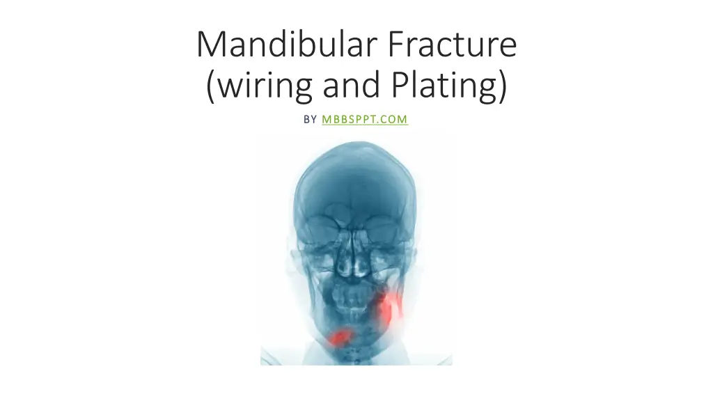
Mandibular Fracture (wiring and Plating) BY BY MBBSPPT.COM MBBSPPT.COM
Different Types of Dental Wiring Different Types of Dental Wiring Techniques Techniques Essig s wiring Gilmer s wiring Risdon s wiring Ivy eyelet wiring
Armamentarium for Wiring Armamentarium for Wiring Presterilized 26-G stainless steel wire Two needle holders or wire holders. Wire cutters.
Essigs wiring Essig s wiring Used to stabilize dentoalveolar fractures. Steps: The luxated teeth should be pushed back into their sockets. The wire is passed around the neck or the chosen teeth, One end going from buccal to lingual and other end going lingual to buccal in each interdental space of teeth And the wires are brought together and twisted and cut short to be tucked into interdental space. The additional small wires are passed interdentally around these base wires to secure them tightly beyond the cingulum. The individual interdental wires are also twisted, cut and adjusted in the interdental spaces.
Gilmers Wiring Gilmer s Wiring It is direct wiring method of intermaxillary fixation. At least one tooth anterior and one posterior to the fracture should be available for wiring to assure proper stabilization. Steps : Stainless steel wire is passed around the neck of the chosen tooth. Both the ends are brought out on the buccal surface and manually twisted keeping the twists close to the tooth. Final twisting is completed by grasping both the ends with a wire holder.
Couple of teeth are chosen in the individual arches and the twisted wires are left long clamped. After the reduction of the fracture is carried out, then the mandibular wires are twisted tightly together with their corresponding maxillary wires. The finally twisted wires are then cut short and the sharp ends turned into the interdental space.
Risdons Wiring Risdon s Wiring Commonly utilized method of horizontal wire fixation. In this method usually second molars on either side are chosen for anchorage. 26-G, 25 cm long wire is passed around the neck of the second molar on each side and both the ends are brought out on the buccal side. Then the ends of both wires twisted together for their entire length, so that the strong base wire is formed on either side, coming towards the midline from each second molar.
The excess wire is cut and the ends tucked in the interdental space.. The base wire is secured to individual tooth by using additional interdental wires. Here small wires are cut and one end of the wire is passed from the distal surface of the tooth below the base wire and brought out towards the lingual side and then brought out on the buccal surface from the mesial interdental space above the base wire. Both these ends are again grasped together and twisted, cut and finished in the interdental space. Each tooth is engaged in the same manner to the base wire, so that the base wire is fully secured to the dental arch.
Ivy Eyelet Wiring Ivy Eyelet Wiring The Ivy loop embraces the two adjacent teeth. 26-G stainless steel wires are used. loop is formed in the center of the wire. The two tail ends of the eyelet are passed through the interdental space of the selected two teeth from buccal to lingual side. One end is passed around distal tooth from lingually to buccally. Other end is passed around mesial tooth lingually to buccally. Twist both the ends and cut it short.
Mandibular fracture wiring Mandibular fracture wiring Transosseous Wiring (Intraosseous Wiring) Intraoral Transalveolar or Upper Border (Superior Border) Wiring Extraoral Lower Border (Inferior Border) Transosseous Wiring
Transosseous TransosseousWiring (Intraosseous Wiring (Intraosseous Wiring) Wiring) Can be done through intraoral or extraoral approach. In principle, holes are drilled in the bony fragments on either side of the fracture line, After which a length of 26-g stainless steel wire is passed through the holes and across the fracture. After this initial preparation, the fracture must be reduced independently with the teeth in occlusion before the free ends of the wire are lightened and twisted. The twisted ends are cut short and tucked into the nearest drill hole. The single strand wire fixation in this horizontal manner is the simplest form of fixation with intraosseous wiring The variations can be: two-hole, four-hole, three-hole technique. Obwegeser s figure-of-eight wiring, Hayton-William s modification of figure-of-eight wiring.
These variations are mainly used at the inferior border of the mandible through extraoral incision.
Intraoral Intraoral Transalveolar Transalveolaror Upper Border (Superior Border) Wiring (Superior Border) Wiring or Upper Border To control the posterior fragment by drilling a hole through the alveolar process of each fragment. Here, the wire is passed through the extraction socket of the third molar tooth, which is invariably involved in the fracture line. Many times, simple loop through only the buccal plate may be adequate. But for better fixation both buccal as well as lingual plates of the alveolar process should be involved and the horizontal mattress type of wiring provides optimum stability. While drilling the holes on either side of the fractured fragments at the alveolar crest, one has to select the site for drilling the holes carefully, so that during final twisting, the thin alveolar bone should not get crumbled down. While drilling the hole on the lingual plate, protection of the lingual nerve should be done by placing periosteal elevator.
Extraoral Lower Border (Inferior Border) Extraoral Lower Border (Inferior Border) Transosseous Transosseous Wiring Wiring The entire area should be well exposed by using L-shaped retractors at the upper edge of the incision. The holes should be drilled using small round bur with electrical engine and handpiece with constant irrigation of saline solution.
The first hole should be drilled in the anterior fragment slightly away from the inferior border (No. 1) and at least 0.5 cm from the fracture site. The bur hole should be through and through the buccal and lingual cortex. Care should be taken that the hole should be drilled away from inferior alveolar nerve. If four-hole technique is to be used, then another hole (No. 2) is drilled above the first hole in the anterior fragment. One hole is placed near the inferior border(No. 3) 0.5 cm from the fracture site. Another hole (No. 4) is placed as high as possible above the first one and just below the inferior alveolar canal. Two separate double wires of 26-G are cut and one wire is passed from No. 1 hole from buccal cortex to lingual cortex and passed at the posterior fragment to lingual cortex and brought out over buccal cortex from hole No. 4. Then another wire is passed from hole No. 2 and brought out from hole No. 3. Both these wire s ends are twisted individually in crisscross manner after approximating the fragments and ends cut and finished.
Bone Plating In Mandibular Bone Plating In Mandibular Fractures Fractures Advantages : Rigid or stable fixation Removes the need for immobilization of the mandible. Soft diet can be taken Maintenance of oral hygiene Useful in mentally challenged, physically handicapped patients Maintenance of airway in multiple fractures.
Simple Simple Noncompression Noncompression Bone Plates Bone Plates Made up of stainless steel or titanium. Screws 2 mm in diameter and 10 mm in length various sizes and shapes.
Miniplate Osteosynthesis ( Miniplate Osteosynthesis (Noncompression Monocortical Monocortical Screw System) Screw System) Noncompression Aim: To attain a functionally oriented fracture adaptation. Fixation with miniplate system, which is as small as possible. Application of the plate to the region of the traction side of the bone. Champy s Ideal Osteosynthesis Lines : Mandible has parabola-shaped body . It consists of the outer and inner cortical plates with central spongiosa. The external cortex is strong and thicker in chin region.
In every mandibular fracture, the forces of mastication produce tension forces at the upper border and compression forces at the lower border. Therefore, distraction of the fractured fragments can be seen at the alveolar crest region. In the canine region, there are overlapping tensile and compressive loads in both the directions.
The determination of these forces allows the establishment of ideal osteosynthesis lines for the mandibular body. It corresponds to the course of a line of tension at the base of the alveolar process. In this region a plate can be fixed with monocortical self-tapping screws. Behind the mental foramen, a plate can be applied immediately below the root apices and above the inferior alveolar nerve. At the angle of the jaw, the plate is most favorably placed on the broad surface of the external oblique line as high as possible. In the anterior region between the mental foramina, in addition to the subapical plate, another plate near the lower border of the mandible is necessary in order to neutralize torsional forces. Second plate is applied parallel to the first plate with a gap of 4.5 mm between them
Miniplate Osteosynthesis Miniplate Osteosynthesis Application of miniplate osteosynthesis the approach is always intraoral, In multiple mandibular fractures, the precise reduction and the establishment of occlusion is more difficult, hence it is advisable to secure the occlusion by interdental wiring, before the osteosynthesis is performed. In the edentulous jaw, manual reduction is generally sufficient.
The adaptation of the bone plate should be carried out by using bending pliers.
The adapted bone plate should lie passively on the contour of the external cortex, without any gap between the plate and the bone. The plate is adjusted in such a way that minimum two screws can be fixed on either side of the fracture line. The drilling is then performed through the holes in the plate perpendicular to the surface of the bone. The miniplate is fixed in position with the screws.
Surgical Procedure of Surgical Procedure of Minibone And Screw And Screw Minibone Plates Plates
Complications and Management Complications and Management Suture dehiscence: It is found if the surgery for osteosynthesis is delayed due to some reasons: Timing of surgery. Due to inappropriate incision in the region of the attached gingiva, on 4th or 8th postoperative day, the gap will be noticed. In such a case, the sutures are removed and cleaning of the wound with 15% hydrogen peroxide is done. Frequent mouthwashes are encouraged. Wound is allowed to heal with secondary intention.
Compression Plates Compression Plates Whenever the plate is applied to the convex surface of the mandible at its lower border, there is a tendency for the upper border and lingual plate to open up with the final tightening of the screws. This leads to distortion of occlusion as well as opening of the fracture line on the other side, in case of bilateral fractures. To overcome this problem and to produce more rigid fixation various designs of compression plates.
These compression plates include at least two-pear shaped holes. The widest diameter of the hole lies near the fracture line. The screw is inserted in the narrow part of the hole and at the final movement of tightening, its head comes to rest in the widest diameter of the hole. The compression holes in the plate may be positioned one on each side of the fracture line.
Reconstruction Plates Reconstruction Plates These are capable of temporary load bearing and therefore useful in comminuted fractures, defect fractures and infected fractures. It is also useful in the case of severely displaced angle fractures. Reconstruction plate absorbs all functional loads to restore the continuity and permits early mobilization, despite extensive fragmentation of the bone. It also spans the comminuted area to preserve the original shape, length and strength of the mandibular arch.
Reconstruction plates must be accurately contoured to avoid fracture displacement and a subsequent malocclusion during tightening of the screws. The use of template simplifies the bending process of the large reconstruction plates. When using a reconstruction plate, the most posterior screw holes to the fracture site, should be placed at least 1 cm away from the fragment end and four screws in each segment provide the maximum resistance to deformation
Complications of mandibular fracture Complications of mandibular fracture management management Infection Bleeding Lip numbness Malocclusion Nonunion Malunion Trismus Tooth loss Paresis Cosmetic compromise are reported to range as high as 40% or more.


")