Osteoarthritis is a common joint disease affecting the articular cartilage, subchondral bone, and synovium. This condition predominantly affects weight-bearing joints like knees and hips, with a higher prevalence in women and older individuals. Learn about the epidemiology, joint classification, and anatomy of synovial joints in osteoarthritis.
Please find below an Image/Link to download the presentation.
The content on the website is provided AS IS for your information and personal use only. It may not be sold, licensed, or shared on other websites without obtaining consent from the author. If you encounter any issues during the download, it is possible that the publisher has removed the file from their server.
You are allowed to download the files provided on this website for personal or commercial use, subject to the condition that they are used lawfully. All files are the property of their respective owners.
The content on the website is provided AS IS for your information and personal use only. It may not be sold, licensed, or shared on other websites without obtaining consent from the author.
OA - Definition Heterogeneous group of conditions resulting in common histopathologic and radiologic changesinvolving Entire joint organ, including: the articularcartilage the subchondral bone and the synovium.
Epidemiology Internationally, osteoarthritis isthe most common articular disease. Estimates of itsfrequency vary across different populations. 80-90%of individuals older than 65 years have evidence of radiographic osteoarthritis. the prevalence of osteoarthritis ishigher among women than among men. Interethnic differences in the prevalence of osteoarthritis have been noted.
Involved joints Weight-bearing joints,including: the knees the hips cervical andlumbosacral spine feet. Non weight bearingjoints: the(DIP), the(PIP), and the(CMC) joints.
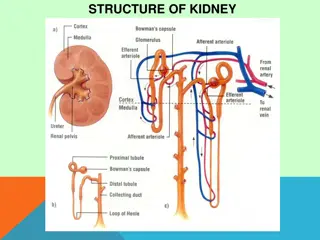
The normal articular surface of synovialjoints articular cartilage (chondrocytes) surrounded by extracellular matrix includes proteoglycans and collagen. The cartilage facilitates joint function and protects the underlying subchondral bone by distributing large loads, maintaining low contactstresses, and reducing friction atthe joint.
Synovial Fluid Synovial fluid is formed by(synoviocytes). Synovial cells also manufacture hyaluronic acid (HA, also known as hyaluronate), a glycosaminoglycan that is the major noncellular component of synovialfluid. Synovial fluid supplies nutrients to the avascular articular cartilage; it also provides the viscosity needed to absorb shock from slow movements provides elasticity required to absorb shock from rapid movements.
There is Inflammation inOA Inflammation occurs as cytokines and metalloproteinases are released into the joint. These agents are involved in the excessive matrix degradation that characterizes cartilage degeneration in osteoarthritis.
Pathogenesis Swelling of the cartilageusually occurs the level of proteoglycans eventually drops very low, the cartilage softens and lose elasticity and compromising joint surface integrity. Flaking and fibrillations (vertical clefts) develop along on the surface of an osteoarthritic joint. Over time, the loss of cartilage results in loss of joint space. a greater lossof joint space occurs at those areas experiencing the highest loads.
Bone changes Bone denuded of itsprotective cartilage continues to articulate with the opposing surface. Eventually, the increasing stresses exceed the biomechanical yield strength of thebone. The subchondral bone responds with vascular invasion and increased cellularity, becoming thickened and dense (a process known as eburnation) at areas ofpressure.
Bone changes subchondral boneundergo cystic degeneration. Osteoarthritic cysts are also referred to as subchondral cysts, pseudocysts, or geodes and may range from 2to 20 mm in diameter. Osteoarthritic cysts in the acetabulum are termed Egger cysts.
Joint changes vascularization of subchondralmarrow, osseous metaplasia of synovial connective tissue, and ossifying cartilaginous protrusions lead to irregular outgrowth of new bone (osteophytes). Fragmentation of these osteophytes or of the articular cartilage itself results in the presence of intra-articular loose bodies (joint mice).
Osteoarthritis Progression Stage 1-breakdown of the cartilage matrix occurs. Stage 2- involves the fibrillation and erosion of the cartilage surface Stage 3-a chronic inflammatory response in the synovium. Further Progression -the above events alter the joint architecture, compensatory bone overgrowth occurs. joint architecture is changed mechanical and inflammatory stress occurs on the articular surfaces, the disease progressesunchecked.
Primary Osteoarthritis Three subtypes Primary generalized OA Erosiveosteoarthritis Chondromalacia Patellae
Work Up Laboratory Plain Radiography CT scan, MRI scan,ultrasonography Bone scintigraphy Arthrocentesis
Treatment Non pharmacologic- Life style modification, physical and rehab therapy Pharmacotherapy Arthroscopy Osteotomy Arthroplasty Fusion and jointLavage Stem cell therapy
References 1.1.Hunter, W. Of the structure and diseases of articulating cartilages. Phil. Trans. Royal Soc. 470, 514 521(1743). 2.2.National Collaborating Centre for Chronic Conditions(UK). 3.National Clinical Guideline for Care and Management in Adults(Royal College of Physicians of London, 2008). 3.3.Felson, D. T. An update on the pathogenesis and epidemiology of osteoarthritis. Radiol. Clin. North Am. 42, 1 9(2004). 4.4.WHO. The World Health Report 2002: reducing risks, promoting healthy life (WHO, 2002). 5.5.Centers for Disease Control and Prevention (CDC). Prevalence of doctor-diagnosed arthritisand arthritis-attributable activity limitation United States, 2010 2012. MMWR Morb. Mortal. Wkly Rep. 62, 869 873 (2013).