Osteomalacia in Adults: Causes, Symptoms & Management
Osteomalacia, also known as adult rickets, is a condition characterized by softening of bones due to a lack of vitamin D. Learn about its causes, symptoms, diagnostic investigations, and management strategies, including dietary recommendations and surgical options.

Download Presentation

Please find below an Image/Link to download the presentation.
The content on the website is provided AS IS for your information and personal use only. It may not be sold, licensed, or shared on other websites without obtaining consent from the author. If you encounter any issues during the download, it is possible that the publisher has removed the file from their server.
You are allowed to download the files provided on this website for personal or commercial use, subject to the condition that they are used lawfully. All files are the property of their respective owners.
The content on the website is provided AS IS for your information and personal use only. It may not be sold, licensed, or shared on other websites without obtaining consent from the author.
E N D
Presentation Transcript
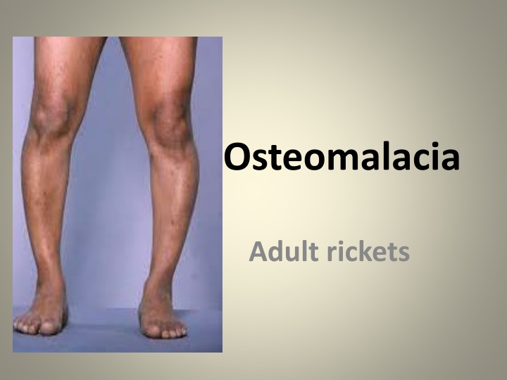
Osteomalacia Adult rickets
Word Meaning Greek origin Osteo bone Malacia - softness
Incidence Women mostly affected (Muslims) Its endemic in Asia
Lack of exposure to UV rays (needed for Vit.D synthesis) Drugs anticonvulsants (phenytoin) GI malabsorption Extensive burns Chronic diarrhoea Pregnancy multiple pregnancy Kidney disease Kidney disease
Excessive loss of calcium (celiac disease, biliary tract obstruction, chronic pancreatitis) After surgery of gastrointestinal system
Pathophysiology Vit.D insufficiency Vit.D required for the absorption of the calcium from the intestine Insufficient vit.D intake can interfere with the normal mineralization of bone activity or insufficient calcification of bone Leads to bone softening and associated symptoms
Clinical features Bone become bent and flattened as they soften Pathologic fracture Fatigue, malaise, bone pain (with tenderness) Difficult to walk, rise from a chair, low back pain, progressive muscle weakness, weight loss , deformity of spine (kyphosis), fracture with delayed healing
Diagnostic investigation X-ray generalized demineralization with trabecular bone loss Bone biopsy Serum calcium and phosphorus levels are reduced Alkaline phosphates level is moderately elevated
Management Diet vit D, calcium and phosphorous rich Vit.D3 (cholecalciferol), vit.D2 (ergocalciferol) Calcium salts and phosphorous supplements may also prescribed Eggs, low fat milk, fish Exposure to sunlight Weight bearing exercises Orthopedic deformities may be treated with surgery (osteotomy).
Nursing management Assessment Assess for bone pain in the low back and extremities Assess for fracture Obtain information about co existing diseases (malabsorption syndrome) and dietary habits Note skeletal deformities on physical examination
Relieving pain change in positions and analgesic Improving body image trusting relationship, encourage patient to discuss any changes in body image and methods of coping Encourage to use existing strength Allow talk with the similar successful patients
Nsg diagnosis Acute pain r/d to bone tenderness and possible fracture Disturbed body image related to bowing of legs, waddling gait and spinal deformities Deficient knowledge about disease process and treatment
Def - idiopathic Skeletal bone disorder in which there is excessive bone resorption followed by replacement of altered bone tissue The new bone is larger, disorganized and structurally weaker Common sites pelvis , long bones, spine, ribs, sternum and cranium
Etiology Exact cause is unknown Followed by viral infection (measles virus, respiratory syncytial virus ) Genetic (15%-30%) Men to women ratio 2:1 Seen in below 40 years
Pathophysiology Due to unknown causes Increase BONE RESORPTION Altered BONE TURN OVER As a compensatory mechanism - increase bone formation Newly formed bones are large, unorganized and weak Structural changes in weight bearing limbs and other manifestation
Signs and symptoms Free of symptoms in the early stages Bone pain deep ache progress to severe pain Fatigue Progressive development of waddling gait Become shorter Barrel chest
Bowing of tibia or femur Changes in skin temperature Manifestations related to nerve compression
Increase bone volume in the spine can cause spinal cord or cranial root compression leads to head ache, blindness, vertigo, hearing loss with tinnitus and dementia Pathologic fracture , osteosarcoma, fibrosacroma and osteoclastoma tumors are complications Osteoarthritis are common associated with pagets disease
Diagnostic investigation X rays shows increased bone expansion and density. Bone scan Serum alkaline phosphates levels increased N-telopeptide and C-telopeptide they are biomarkers used to measure the rate of bone turnover, can be measure from urine or serum
Management Limited to symptoms Supportive care Correction of secondary deformity Calcitonin decrease osteoclastic activity
Bisphosphonate drugs Risedronate, Tidronate, Pamidronate, Ibandronate retard bone resorption Calcium and vitamin D supplements Pain NSAIDS, ibuprofen. Orthopedic surgeries for fracture, hip and knee replacements Firm mattress back support and to relieve pain Wear light brace Activity like lifting and twisting should be avoided
Physical therapy will increase muscle strength Good body mechanics Balanced nutrition programme vit.D, calcium and protein Massage, heat therapy Prevention Patient education Use of an assistive device


")