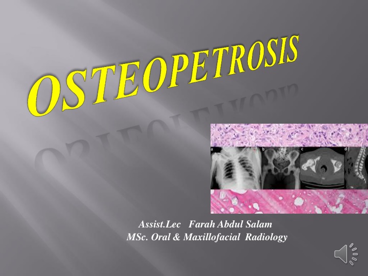
Osteopetrosis: Characteristics, Mechanisms, and Management
Learn about osteopetrosis, a hereditary skeletal disorder characterized by increased bone density due to abnormal osteoclast function. Discover the history, clinical features, mutations, primary mechanism, clinical variants, therapeutic options, supportive measures, lab findings, skeletal X-ray findings, and more related to this condition.
Download Presentation

Please find below an Image/Link to download the presentation.
The content on the website is provided AS IS for your information and personal use only. It may not be sold, licensed, or shared on other websites without obtaining consent from the author. If you encounter any issues during the download, it is possible that the publisher has removed the file from their server.
You are allowed to download the files provided on this website for personal or commercial use, subject to the condition that they are used lawfully. All files are the property of their respective owners.
The content on the website is provided AS IS for your information and personal use only. It may not be sold, licensed, or shared on other websites without obtaining consent from the author.
E N D
Presentation Transcript
Assist.Lec Farah Abdul Salam MSc. Oral & Maxillofacial Radiology
History Definition Clinical Features Mutations associated with it Primary Mechanism involved It s Clinical Variants Therapeutic Options Supportive Measures Lab. Findings Skeletal X-ray Findings Conclusion
Osteopetrosis Disease History Osteopetrosis was first described , by a German radiologist Albers-Sch nberg
A Hereditary skeletal disorder characterized by marked increase in bonedensity Bone density is due to defect in remodeling caused by Abnormal Osteoclast Function Defective osteoclast resorption withcontinued bone formation and endochondral ossification thickening of cortical bone and cancellous bonesclerosis results
Fractures Low Blood Cell Production Loss of Cranial Nerves Function Frequent Infections of Teeth & jaws
:H+ protonpump Carbonic anhydraseII -ATP ase
a deficiency of the carbonic anhydrase enzyme encoded by the CA2 gene. Carbonic anhydrase is required by osteoclasts for proton production. Without this enzyme hydrogen ion pumping is inhibited and bone resorption by osteoclasts is defective, as an acidic environment is needed to dissociate calcium hydroxyapatite from the bone matrix. As bone resorption fails while bone formation continues, excessive bone is Formed.
INFANTILE i) malignant ii)intermediate iii) transient ADULTOSTEOPETROSIS
Severe disease at birth or early infancy MALIGNANT It is autosomal recessive Diffusely scleroticskeleton Initial signs Normocytic anemia withhepatosplenomegaly extramedullaryhematopoiesis) granulocytopenia) Increase Susceptibility to infection
Facial deformity(broad face, hypertelorism,sunb nose Delayed tooth eruption Narrowing of skull foramina (failure of resorption and remodelling) pressing on cranial nerves optic nerve atrophy,blindness,deafness,facialparalysis. Osteomyelitis jaw
Bone in Bone appearance
Less severe variant of infantileosteopetrosis Affected patients asymptomatic atbirth Fractures exhibited by the end of firstdecade Rarely marrowfailure/hepatosplenomegaly
Radiographic evidence of diffusesclerosis Marrowfailure Resolve without specifictherapy Most affected patients return tonormal without sequelae
Less severe manifestations Autosomaldominant trait---benign osteopetrosis Sclerosis of axialskeleton Long bones little/no defect 40% patients asymptomatic marrow failurerare
Plain film radigraphsreveal a diffuse increased radiopacity of medullary portions ofbone Symptomatic patients bonepain Two major variants with CN compression: rarely fractures occur without CN compression: fractures common. mandible osteopetrosis: complications:fractures and osteomyelitis
Other therapies: interferron gama-1b incombination with calcitriol-reduce bone mass,decrease infections, lowers nervecompressions Others: corticosteroid administration(toincrease circulating RBCS and Adult osteopetrosis-mild severity-long termsurvival Infantileosteopetrosis-poor prognosis-most deaths during first decade oflife Bone marrow transplantation onlyhope for cure platelets) Parathormone Macrophagecolony stimulating factor Erythropoietin Limiting calciumintake
Transfusions Antibiotics Osteomyelitis of jaws: drainage,debridement,culture with sensitivity,appropriate antibiotic therapyand reconstruction if necessary
Lateral radiograph of the skull reveals diffuse thickening of the calvarium, most significant in the region of the occiput. The partially visualized upper cervical vertebrae and maxilla are also dense andthickened.
Sagittal T1-weighted MR image shows thickening of the calvaria and facial bones with hypointensity of skull and cervical vertebra, and cerebellar tonsillar ectopia.
Axial T2-weighted image shows optic canal stenosis and optic nerve atrophy
Severe bilateral optic canal narrowing (arrows) in a 4yr-old patient with complete loss of vision in the left eye and 20/80 visual acuity in the right eye.
Spine radiographs reveal the classic sandwich vertebrae of osteopetrosis (red arrows). This is manifested as thickening and sclerosis of the vertebral endplates, and of the bone adjacent to the endplates. There is also marked thickening of the posterior vertebrae (yellow arrows), especially in the vertebral arch.
Chest radiograph obtained in an infant demonstrates overall increased density of the osseous structures due to the accumulation of immature bone.
Generalized increased density of the bones and alternating areas of increased and decreased density in the metaphysis (bone- within-bone appearance).
Densely sclerotic bone ,,,,, deformity of the femurs with under- trabeculation.
Heavy Metal Poisoning e.g. (Lead) Melorheosis Hypervitaminosis D ( Vit.D toxicity) Pyknodysostosis Fibrous Dysplasia of bones
Q/What is the difference between Osteoporosis & Osteopetrosis???
Osteopetrosis is a rare hereditary skeletal disease that can be appeared early in life as (Infantile Congenita) or as an (Adult Osteopetrosis) It s Diagnosis is based on Thorough clinical evaluation, detailed pt. history, with a variety of specialized tests like [[ X-ray imaging,measuring of BMD]] Skeletal X-ray Findings very specific & considered sufficient to make a diagnosis
