OVCA Tumor Drug Transport Proteins and RAB27A Expression Analysis
Explore the correlation between drug transport proteins, exosome-associated proteins, and RAB27A expression in ovarian cancer tumors. Discover the prognostic significance of exosomes and CA125 in ovarian cancer outcomes. Validated biosensor for detecting plasma-derived small EVs and CDDP. Investigate the role of pGSN in regulating CDDP and exosome secretion in HGS ovarian cancer cells.

Uploaded on | 1 Views
Download Presentation

Please find below an Image/Link to download the presentation.
The content on the website is provided AS IS for your information and personal use only. It may not be sold, licensed, or shared on other websites without obtaining consent from the author. If you encounter any issues during the download, it is possible that the publisher has removed the file from their server.
You are allowed to download the files provided on this website for personal or commercial use, subject to the condition that they are used lawfully. All files are the property of their respective owners.
The content on the website is provided AS IS for your information and personal use only. It may not be sold, licensed, or shared on other websites without obtaining consent from the author.
E N D
Presentation Transcript
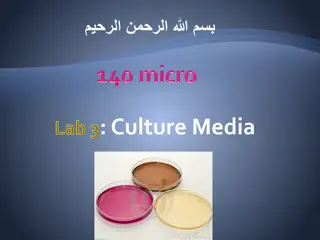
Supp. Fig. 1: OVCA tumor expressions of drug transport proteins and RAB27A A P-gp MRP2 RAB27A P=0.02 P=0.32 P=0.33 AUC: 0.57 P=0.009 AUC: 0.53 P=0.15 AUC: 0.53 P=0.18 C B Exosome-associated protein Drug transport proteins pGSN pGSN RAB27A MRP2 P-gp Supp. Fig. 1. Correlation between pGSN and drug transport proteins as well as exosome-associated proteins in OVCA patient tumors. (A) Our interrogation of TCGA public dataset revealed that P-gp but not MRP2 and RAB27A are significantly predictive of chemoresistance in human OVCA tumors. (B) A positive correlation was observed between pGSN and MRP2, P-gp and (C) RAB27A. N=958
Supp. Fig. 2: pGSN regulates CDDP and exosome secretion in HGS OVCA cells A New.OV90.siRNA.untreated.exosomes Chemoresistant HGS; OV90 B 400 OV90 OV90siRNA Exosomes sEV (nM) 300 (nM) 200 100 0 OV90 OV90siRNA Supp. Fig. 2. pGSN regulates exosomal release in high grade serous (HGS) OVCA cell lines. (A) Human recombinant pGSN was synthesized with Myc and 6His tags inserted. The refolded gel UV absorbance measured with A280=0.0210497. pGSN downregulation in chemoresistant HGS cells reduced sEV production whereas the vice-versa occurs when pGSN chemosensitive HGS cells. pGSN was (B) silenced in chemoresistant cells (siRNA; 50 nM, 24 h) and (C) over-expressed in chemosensitive cells (cDNA 2 ug, 24 h). Small EV concentrations were determined by biosensor-sEV aggregation using Raman spectroscopy. Results are expressed as means SD from three independent replicate experiments. New.TOV3133.pGSN.untreated.exosomes Chemosenistive HGS; TOV3133 New.TOV3041G.pGSN.treated.exosomes Chemosensitive HGS; TOV3041G C 150 150 TOV3133 TOV3133 pGSN 3041G 3041GpGSN Exosomes sEV (nM) Exosomes 100 100 (nM) (nM) in over-expressed in 50 50 0 0 TOV3133 TOV3133 pGSN 3041G 3041GpGSN
Supp. Fig. 3: Validation of biosensor on human OVCA plasma-derived EVs A B 300 100 Predicted concentration (%) Raman Intensity (AU) Rate (cts/min) 150 50 0 + - - - 0 - - Buffer A Buffer B CDDP - + + + 50 0 1000 400 100 + + 1600 Raman Shift (cm-1) Actual concentration (%) Supp. Fig. 3. Plasma-derived small EV isolation, characterization and biosensor detection. (A) Biosensor detection of small EVs and CDDP were performed using buffer A and B from the Exoquick Ultra kit. (B) Raman peaks of Buffer A and B were generated to determine their composition and potential influence on exosome and CDDP detection.
Supp. Fig. 4: Prognostic significance of exosome and Ex/CA125 in OVCA patients A sEV (nM) sEV (nM) sEV (nM) sEV (nM) sEV (nM) sEV (nM) B C sEV (nM) CA125 sEV (nM) CA125 Supp. Fig. 4. Prognostic significance of sEVs and sEVs/CA125 in OVCA outcomes . Small EVs were isolated from the plasma of human OVCA patient with pre-determined CA125. (A) Mean SD of sEV or sEV/CA125 were determined and compared within clinical outcomes (diagnosis, residual disease, chemoresistance, stage, tumor recurrence and CA125 levels). (B) Plasma-derived sEVs were correlated with CA125 in malignant OVCA patients. (C) The mean SD levels of sEV/CA125 were determined and compared within OVCA clinical outcomes (residual disease and histological subtype). sEV (nM)