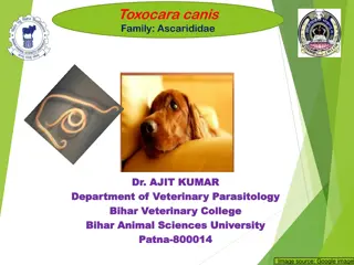
Overview of Ehrlichia Infections in Dogs
Ehrlichia is a genus of bacteria that can cause diseases in dogs, such as canine rickettsiosis. From transmission to pathogenesis, this article explores various aspects of Ehrlichia infections in dogs, including the morphology, definitive hosts, transmission methods, and associated diseases. Learn about the impact on puppies and how stress and concurrent infections can exacerbate the illness.

Download Presentation

Please find below an Image/Link to download the presentation.
The content on the website is provided AS IS for your information and personal use only. It may not be sold, licensed, or shared on other websites without obtaining consent from the author.If you encounter any issues during the download, it is possible that the publisher has removed the file from their server.
You are allowed to download the files provided on this website for personal or commercial use, subject to the condition that they are used lawfully. All files are the property of their respective owners.
The content on the website is provided AS IS for your information and personal use only. It may not be sold, licensed, or shared on other websites without obtaining consent from the author.
E N D
Presentation Transcript
The Diabetic Retinopathy Clinical Research Network Intravitreous Anti-VEGF vs. Vitrectomy for Vitreous Hemorrhage from PDR (Protocol AB) Protocol Chair: Andrew N. Antoszyk, MD
Background Vitreous hemorrhage due to PDR is a frequent cause of vision loss in patients with diabetes. No recent, definitive evidence to guide how to: Optimize VA outcomes, or Optimize time to maximal visual recovery Major question of interest: Once intervention is desired, what is the relative safety and efficacy of currently available therapies? 2
Anti-VEGF for VH in PDR Rationale Data supporting anti-VEGF use for VH + PDR: In DRCR.net Protocol N, a difference in rates of vitrectomy at 4 months with anti-VEGF compared with saline-treated eyes was not identified BUT Ranibizumab treated eyes showed some biological effects suggesting benefits: Higher probability of completing PRP (44% vs 31% by 16 weeks P = 0.05) Greater average VA improvement (22 vs. 16 letters, P = 0.04) Reduced rate of recurrent hemorrhage (6% vs. 17%, P = 0.01) 3
Vitrectomy for VH in PDR Rationale Newer surgical techniques allow smaller incisions, faster operating times, and fewer associated adverse events DRIVE UK study: Eyes with simple VH of at least 3 months duration had better outcomes than eyes with TRD or other diabetic pathology 85% (51) had >3 line gain in VA Mean gain of 42 27 ETDRS letters 8.3% (5) had repeat vitrectomy 3 for non-clearing VH; 2 for RD Gupta B, Sivaprasad S, Wong R, et al. Visual and anatomical outcomes following vitrectomy for complications of diabetic retinopathy: the DRIVE UK study. Eye (Lond). 2012 Apr;26(4):510-6. 4
Study Objectives 1. Evaluate and compare VA outcomes over time for the following regimens: A. Prompt vitrectomy + PRP B. Intravitreous anti-VEGF (Aflibercept) injections 2. Characterize the follow-up course for the two treatment regimens, including: Post-operative complications for the vitrectomy group Number of injections needed and percent requiring vitrectomy in the aflibercept group 5
Study Design Multi-Center Randomized Clinical Trial 200 eyes (1 per pt) that meets the following criteria: Vitreous hemorrhage 1. causing vision impairment, 2. presumed to be from PDR, and 3. requiring intervention (vitrectomy or anti-VEGF). Vitrectomy + PRP Anti-VEGF Primary Outcome: Visual Acuity Area Under the Curve (AUC) over 6 Months* Total Duration: 2 Years 6 *Note: Publication not planned until 2-year results available
Additional Key Outcomes Although 6M VA AUC is primary , long- term outcomes are also important: Mean VA at 1, 3 months and annual visits VA AUC at annual visits Percent 20/20 and 20/40 or better and 20/200 or worse at annual visits Rates of recurrent VH on clinical exam Productivity from WPAI questionnaire Treatment and follow-up costs 7
Major Inclusion/Exclusion Criteria VA 20/32 or worse but at least light perception No anti-VEGF treatment within 2 months prior to vitreous hemorrhage onset No prior vitrectomy Note: Prior PRP is not a requirement nor an exclusion No rhegmatogenous retinal detachment and no traction retinal detachment involving or threatening the fovea Patient is able and willing to undergo vitrectomy within 2 weeks (no contraindication to surgery) 8
Follow-Up and Treatment Overview Outcome assessment visits for both groups Every 3 months in the first year Every 4 months in the second year Anti-VEGF Treatment Visits every 4 weeks for 6 months and then as needed Injections required in the Anti-VEGF Group through week 12 and then as needed according to criteria Both groups may receive anti-VEGF for DME Vitrectomy Within 2 weeks of randomization in the Vitrectomy Group If needed in anti-VEGF group according to criteria 9
Referrals We Need Your Help! Please consider any eyes with vitreous hemorrhage due to diabetic retinopathy causing vision impairment Study participants must be willing to be randomized to vitrectomy or aflibercept injections and continue follow-up for 2 years 10
Thank You on Behalf of Diabetic Retinopathy Clinical Research Network (DRCR.net) Dedicated to multicenter clinical research of diabetic retinopathy, macular edema and associated disorders. 11