Overview of Hernia: Definition, Anatomy, Types, Complications & Treatment
Hernia involves the displacement of tissue between compartments due to pressure. Learn about abdominal wall hernia, surgical anatomy, types, complications, and treatments like surgery. Understand the layers of the abdominal wall, inguinal canal, and more.

Download Presentation

Please find below an Image/Link to download the presentation.
The content on the website is provided AS IS for your information and personal use only. It may not be sold, licensed, or shared on other websites without obtaining consent from the author.If you encounter any issues during the download, it is possible that the publisher has removed the file from their server.
You are allowed to download the files provided on this website for personal or commercial use, subject to the condition that they are used lawfully. All files are the property of their respective owners.
The content on the website is provided AS IS for your information and personal use only. It may not be sold, licensed, or shared on other websites without obtaining consent from the author.
E N D
Presentation Transcript
Prepared by: Abdullah Al Saleh Mohammad Al mazroa Khalid Al Qahtani Supervised by: Dr. Fahad Bamehriz
Objectives : Definition of hernia Surgical anatomy Common types and presentation Complications of hernia Surgical treatment
Objectives : Definition of hernia Surgical anatomy Common types and presentation Complications of hernia Surgical treatment
Definition: Hernia is the physical displacement of tissue from one compartment into another due a pressure gradient across the opening between the chambers. Abdominal wall hernia: protrusion of all or part of any intra abdominal structure through any congenital, acquired or iatrogenic defect.
Objectives : Definition of hernia Surgical anatomy Common types and presentation Complications of hernia Surgical treatment
Objectives : Defenition of hernia Surgical anatomy Common types and presentation Complications of hernia Surgical treatment
Surgical anatomy Layers of abdominal wall: 1)skin 4) Internal Oblique Rectus sheath Cremaster muscle 2)Subcutaneous Tissue: ( Superficial Camper s fascia, Deep Scarpa s fascia) 5) Transversus abdominus Rectus sheath Internal or deep inguinal ring 3) External Oblique Rectus sheath inguinal ligament external spermatic fascia external or superficial inguinal ring 6) Transversalis Fascia Internal spermatic fascia 7) Peritoneum
Cont Inguinal canal: 4cm long From the deep ring to superficial ring Above the inguinal ligament Inferior wall (floor): inguinal ligament Superior wall ( roof) : lower fibers of internal oblique and transversusabdominis Content: Spermatic cord in male Round ligament in the uterus in females Walls: Anterior: aponeurosisof external oblique Posterior wall: fascia transversalis
Cont Inguinal Triangle ( Hesselbach's triangle) It is defined by the following structures: Lateral margin of the rectus sheath (medially) Inferior epigastricvessels (laterally) Inguinal ligament (inferiorly)
Objectives : Definition of hernia Surgical anatomy Common types and presentation Complications of hernia Surgical treatment
Objectives : Definition of hernia Surgical anatomy Common types and presentation Complications of hernia Surgical treatment
Types: There are many types of hernias like: Inguinal Hernia Femoral Hernia ObturatorHernia Umbilical Hernia Incisional Hernia Spigelian Hernia Epigastric Hernia Lumbar Hernia Others ..
Inguinal Hernia 1) Indirect Inguinal Hernia: Most common hernia in both sexes. Congenital in origin It occurs when bowels, omentumor any other intra abdominal organ protrudes through the deep ring within a patent processusvaginalis.
Cont. History: Patient may present with a swelling, pain ,or symptoms of complication. Take history of the swelling (when was it noticed, how did the patient notice it?, disappearance, ) If there is pain take history of the pain , and review GI symptoms. Risk factors (lifting heavy object, chronic cough, constipation, previous surgery, trauma, family history )
Cont. Examination: Standing position. Inspection: Site, Shape, uni or bilateral. Measure the size inspect the skin overlying it. inspect for peristalsis. make the patient cough and inspect for increase in size
Palpation: Change in temperature. Tenderness. Can you get above the swelling? Palpate the pubic tuberculeand locate the site of the swelling. Palpate the testis. What is the consistency? Ask the patient to cough and feel for enlargement. Is it reducible? Deep ring occlusion test.
Percussion: Resonant (indicate bowel) Auscultation : Bowel sounds? General examination for causes ( Respiratory, GI, ) Don t forget to examine the other site!
Inguinal Hernia 2) Direct inguinal hernia: In the (Hesselbach's triangle) Doesn t extend through the scrotum Acquired lesion ( more common in older men)
Cont Differential Diagnosis of inguinal hernia: Femoral hernia. Hydroceleof the cord. Un-descended testis. Lipomaof the cord.
Incisional Hernia Can develop through any incision. Deep wound infection is the most common cause of this hernia. Obesityand number of prior operations may play a role. What is the difference between incisional hernia and recurrent hernia?
Epigastric Hernia Congenital or acquired weakness of the midline linea alba It is more common in men. 20 % are multiple.
Spigelian hernia Herniation through semi lunar line. Seen in Obese patients. Common to have a narrow neck.
Others Littre s hernia: A groin hernia that contains Meckel sdiverticulum is called Littre s hernia. Richter s hernia: Only a portion of the bowl incarcerate or strangulate. Symptoms of bowel obstruction is absent!
Cont. Sliding hernia: when a portion of the wall of the protruding sac is made of some intra abdominal organ
Umbilical hernia It s hernia through the umbilical ring. it contains mostly bowel in neonates or omentum in adults. Among adults, it is three times more common in women than in men; among children, the ratio is roughly equal.
Congenital Common in children but usually closes by the age of 2 years, < 5% persist.
Acquired In adults, associated with intra-abdominal pressure e.g. obesity, heavy lifting, a long history of coughing, or multiple pregnancies. **Through a defect adjacent to umbilicus and NOT through the umbilcal scar itself termed PARA- UMBILICAL
Presentation: *Ahernia is present at the site of the umbilicus (commonly called a navel, or belly button) in the newborn; although sometimes quite large, these hernias tend to resolve without any treatment by around the age of 2-3 years. *Obstruction and strangulation of the hernia is rare because the underlying defect in the abdominal wall is larger than in an inguinal hernia
FEMORAL HERNIA Femoral hernias occur just below the inguinal ligament, when abdominal contents pass through a naturally occurring weakness called the femoral canal. Femoral hernias are a relatively uncommon type, accounting for only 3% of all hernias. While femoral hernias can occur in both males and females, but almost all of them develop in women because of the wider bone structure of the female pelvis. It has a narrow neck, 30%-40% of them get incarcerated or strangulated . Risk factors: female, prior pregnancy, prior inguinal hernia repair.
FEMORAL CANAL ANATOMY: Ant: inguinal ligament. Post: cooper s ligament. Med: lacunar ligament. Lat: femoral vein
obturator hernia An obturatorhernia is a rare type of abdominal wall hernia in which abdominal content protrudes through the obturator foramen. it is much more common in women than in men, especially multiparous and older women who have recently lost a lot of weight.
presentation Usual presentation is small bowel obstruction of unknown cause. May compress the obturator nerve and cause pain or paresthesia in the medial thigh DX: The diagnosis is often made intraoperativelyafter presenting with bowel obstruction C.T scan The Howship-Romberg sign is suggestive of an obturator hernia, exacerbated by thigh extension, medial rotation and abduction.
Lumbar Hernia In the lumbar region, in the form of a broad bulging hernia, that are not vulnerable to incarceration. Petit s hernia: in the inferior lumbar triangle. Grynfeltt s Hernia: in the superior lumbar triangle and is less common than Petit s.
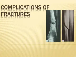
Objectives : Defenition of hernia Surgical anatomy Common types and presentation Complications of hernia Surgical treatment
Objectives : Defenition of hernia Surgical anatomy Common types and presentation Complications of hernia Surgical treatment
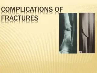
Complication Strangulation Obstruction Incarceration
Reducible: hernial contents can be pushed back into their usual anatomical site in the abdomen. Incarcerated: (imprisoned) hernial contents can NOT be pushed back = irreducible. Strangulated: (choked) the tissue contained in the hernia is ischemic and necrosed due to tocompromise of its blood supply. Obstructing: the hernia contains a loop of bowel that is kinked and obstructs the GI tract. Obstruction is Independent to strangulation.
Incarceration In case of incarcerated hernia: Cannot be reduced (either spontaneously or manually) . Painful enlargement of a previous hernia. Nausea, vomiting, and symptoms of bowel obstruction (possible). An incarcerated hernia could be strangulated, obstructed or both or NONE Every strangulated or obstructed should be incarcerated Reduce it by analgesia, squeeze it by 2 hands to relief edema
Strangulation In case of strangulated hernia:. Symptoms of an incarcerated hernia present combined with a toxic appearance. Strangulation is probable if pain and tenderness of an incarcerated hernia persist after reduction.
Cont For strangulated hernias, start broad-spectrum antibiotics. Antibiotics are administered routinely if ischemic bowel is suspected. Correction of volume status and electrolyte abnormalities. If the pt have strangulated hernia take him to the OR
Obstruction If the pt have obstruction treat him conservatively NPO for 24 hrs If it did not get relived take him to the OR