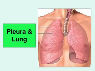
Overview of Lung Anatomy and Features
Lungs are vital respiratory organs located in the thoracic cavity. Each lung has specific characteristics such as conical shape, apex, base, borders, and surfaces. The right lung is heavier than the left, and both undergo color changes with age. Understanding the anatomy, borders, and surfaces of lungs is essential for medical professionals and students.

Download Presentation

Please find below an Image/Link to download the presentation.
The content on the website is provided AS IS for your information and personal use only. It may not be sold, licensed, or shared on other websites without obtaining consent from the author.If you encounter any issues during the download, it is possible that the publisher has removed the file from their server.
You are allowed to download the files provided on this website for personal or commercial use, subject to the condition that they are used lawfully. All files are the property of their respective owners.
The content on the website is provided AS IS for your information and personal use only. It may not be sold, licensed, or shared on other websites without obtaining consent from the author.
E N D
Presentation Transcript
LUNG ANATOMY BY MBBSPPT.COM
INTRODUCTION 1. Lungs are a pair of respiratory organs situated in the thoracic cavity. 2. Each lung innervates the corresponding pleural cavity 3. In the young ,lungs are brown or grey in color. Gradually, they become mottled black because of the deposition of inhaled carbon particles 4. The right lung weighs about 700g.It is about 50- 100g heavier than the left lung.
FEATURES 1. Each lung is conical in shape 2. It has A. Apex at the upper end B. Base resting on the diaphragm C. Three borders, i.e., anterior, posterior, and inferior D. Two surfaces, i.e., costal and medial
1. The apex lies above the level of first rib.It reaches 2.5 cm above the medial one third of clavicle, just medial to supraclavicular fossa. 2. The base rests on the diaphragm which separates the right lung from the right lobe of the liver and the left lung from the left lobe of the liver,fundus of stomach and the spleen 3. The anterior border of the left lung shows a wide cardiac notch below the level of the fourth costal cartilage. The heart and pericardium are uncovered by the lung in the region of this notch.
4. The posterior border corresponds to the medial margins of the heads of the ribs. It extends from the level of 7th cervical spine to the 10th thoracic spine. 5. Inferior border separates the base from costal and medial surfaces. 6. The costal surface is large and convex. It is in contact with the costal pleura and the overlying thoracic wall. 7. The medial surface is divided into a vertebral part and mediastinal part. The mediastinal part shows a cardiac impression, the hilum
FISSURES AND LOBES OF THE LUNGS The right lung is divided into three lobes by two fissures, oblique and horizontal, the left lung is divided into two lobes by oblique fissure
1. The oblique fissure cuts into whole thickness of the lung, except at the hilum Due to the oblique plane of the fissure, the lower lobe is more posterior and the upper and the middle lobe more anterior In the right lung, the horizontal fissure passes from the anterior border upto the oblique fissure and separates a wedge shaped middle lobe from the upper lobe. 2. 3.
1. The tongue shaped projection of the left lung below the cardiac notch is called lingula. It corresponds to middle lobe of the right lung 2. The lungs expand maximally in the inferior direction because movements of the thoracic wall and diaphragm are maximal toward the base of the lung 3. The presence of the the oblique fissure of the each lung allows a more uniform expansion of the whole lung
ROOT OF THE LUNG 1. The root of the lung is a short ,broad pedicle which connects the medial surface of the lung to the mediastinum 2. It is formed by the structures which either enter or come out of the lung at the hilum 3. The roots of lungs lie opposite the bodies of fifth, sixth and seventh thoracic vertebrae
CONTENTS OF ROOT OF THE LUNG 1. Principal bronchus on the left side, and eparterial and hyparterial bronchi on right side 2. One pulmonary artery 3. Two pulmonary veins, superior and inferior 4. Bronchial arteries, one on the right side and two on the left side 5. Bronchial veins 6. Anterior and posterior pulmonary plexuses of nerves 7. Lymphatics of lung 8. Bronchopulmonary lymph nodes 9. Areolar tissue
BRONCHO PULMONARY SEGMENTS 1. These are well defined sectors of the lung each one of which is aerated by a tertiary or segmental bronchus. 2. Each segment is pyramidal in shape with its apex directed towards the root of the lung. 3. These bronchopulmonary segments are independent respiratory units
1. The branches of the pulmonary artery accompany the bronchi, the artery lies dorsolateral to the bronchus, thus each segment has its own separate artery. 2. The pulmonary veins do not accompany the bronchi or the pulmonary artery, they run in inter segmental planes. 3. Thus, each segment has more than one vein and each vein drains more than one segment.
1. Usually the infection of a segment remains restricted to it although some infections like tuberculosis may spread from one segment to the other. 2. However, the segments are no barriers to the spread of bronchogenic carcinoma.
ARTERIAL SUPPLY 1. On the right side there is one bronchial artery which arises from either the third posterior intercostal artery or from the upper left bronchial artery 2. On the left side there are two bronchial arteries both of which arise from the descending thoracic aorta 3. There are pre capillary anastomoses between bronchial and pulmonary arteries. these connections enlarge when any one of them is obstructed in disease
VENOUS DRAINAGE 1. Usually there are two bronchial veins on each side, the right bronchial vein drain into the azygous vein. 2. The left bronchial vein drains either into the left superior intercostal vein or into the hemi azygous vein. 3. The greater part of the venous blood from the lung is drained by the pulmonary veins.
NERVE SUPPLY 1. Para sympathetic nerves are derived from the vagus. These fibres are: motor to the bronchial muscles and on stimulation cause bronchospasm secretomotor to the mucous glands of the bronchial tree the sensory fibres are responsible for stretch reflex of the lungs and for the cough reflex
2. The sympathetic nerves are derived from second to fifth spinal segments. These are inhibitory to the smooth muscles and glands of bronchial tree. 3. This is how sympathomimetic drugs like adrenaline cause brochodilatation and relieve symptoms of bronchial asthma.