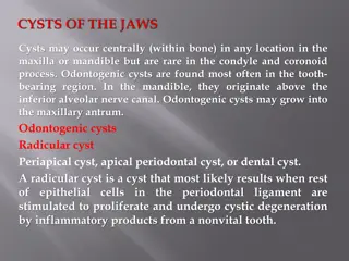
Odontogenic tumors are a group of rare growths that originate from the cells involved in tooth development. These tumors can occur in the jaw bones, gums, or other areas of the oral cavity. Understanding the causes, symptoms, and treatment options for odontogenic tumors is crucial for timely diagnosis and management. While some odontogenic tumors are benign, others can be malignant, highlighting the importance of proper evaluation and treatment by dental professionals. This article provides a comprehensive overview of odontogenic tumors, including their classification, clinical presentation, diagnostic approach, and treatment modalities.
Please find below an Image/Link to download the presentation.
The content on the website is provided AS IS for your information and personal use only. It may not be sold, licensed, or shared on other websites without obtaining consent from the author. If you encounter any issues during the download, it is possible that the publisher has removed the file from their server.
You are allowed to download the files provided on this website for personal or commercial use, subject to the condition that they are used lawfully. All files are the property of their respective owners.
The content on the website is provided AS IS for your information and personal use only. It may not be sold, licensed, or shared on other websites without obtaining consent from the author.
OVERVIEW General consideration Classification Description of some common and important odontogenic tumors.
Odontogenic tumors comprise a complex group of lesions with varied histopathological and clinical features. Some tumors are true neoplasms, while some are hamartomas (developmental malformations). Some are composed only of odontogenic epithelium, while many are mixed i.e. both epithelium and mesenchyme, while some are composed only of mesenchyme
Odontogenic tumors like normal odontogenesis demonstrate varying inductive interactions between odontogenic epithelium & odontogenic ectomesenchyme
Most common odontogenic neoplasm Slowly growing, locally aggressive, benign neoplasm. Occurs in 3 different types with differing clinical, radiological and histological features. 1. CONVENTIONAL SOLID/ MULTICYSTIC 2. UNICYSTIC 3. PERIPHERAL
CONVENTIONAL-AMELOBLASTOMA CLINICAL FEATURES: Age incidence: 3rd-7thdecades. Sex incidence: none Site predilection: 80-85% mandible (posterior) 20%maxillary molar region.
Signs & symptoms Slowly growing, painless, hard bony swelling or expansion of jaw. paresthesia & pain are rare
RADIOLOGICAL FEATURES: Typically rounded, well defined multilocular radiolucency with scalloped margins. When loculations are large, the appearance is called as SOAP BUBBLE appearance.When loculations are smaller, the appearance is called HONEY COMBED appearance.
Desmoplastic ameloblastoma Most common in the anterior regions of the jaws especially the maxilla Mixed lucent & opaque appearance What is the histopathology of the opaque material? osseous metaplasia within the dense fibrous septa NOT a mineralized product DD:fibro-osseous lesions
HISTOPATHOLOGICAL FEATURES: Many subtypes are seen. 1. FOLLICULAR(most common) 2. PLEXIFORM 3. ACANTHOMATOUS 4. GRANULAR CELL 5. DESMOPLASTIC 6. BASAL CELL TYPE What is their significance? none, no effect on their behavior
AMELOBLASTOMA (FOLLICULAR) Islands of epithelium resemble dental organ surrounded by mature connective stroma. Individual follicles show central mass of stellate reticulum like cells surrounded by a single peripheral layer of ameloblast like cells. Nuclei of peripheral cells are reversely polarized. Within the islands, cyst formation is common.
AMELOBLASTOMA (PLEXIFORM) long, anastomosing cords and occasional sheets of epithelial cells bounded by columnar / cuboidal cells. Cells within cords are more loosely arranged than peripheral cells. Supporting stroma is loose and vascular. Cyst formation occurs, not inside follicles, but in surrounding stroma.
AMELOBLASTOMA (ACANTHOMATOUS) Central area of follicles show extensive squamous metaplasia, often associated with keratin formation. DOEAS NOT INDICATE A MORE AGGRESSIVE COURSE OF TUMOR. Can be confused with ??.
AMELOBLASTOMA (GRANULAR CELL) cells show granular cell change. These cells have abundant cytoplasm filled with eosinophilic granules. Seen in younger persons and appears to be more aggressive clinically.
AMELOBLASTOMA (DESMOPLASTIC) small islands / cords of odontogenic epithelial cells surrounded by a dense, collagenized stroma. Peripheral ameloblast like cells are inconspicuous TGF-B
AMELOBLASTOMA (BASAL CELL) Least common type. Composed of nests / sheets of hyperchromatic basaloid cells. No stellate reticulum present centrally and peripheral cells tend to be cuboidal rather than tall columnar.
TREATMENT: Can vary from simple enucleation to curettage to en bloc resection. As lesion spreads through medullary spaces, simple enucleation can leave islands of tumor within the jaws, leading to recurrence. Marginal resection is the optimal method. Post. Maxilla Radio-therapy ??? Rarely can undergo malignant transformation.
UNICYSTIC AMELOBLASTOMA Controversy, whether it arises de novo or as neoplastic transformation of odontogenic cyst lining.
CLINICAL FEATURES: Age incidence: Younger individuals(23y) Site predilection: 90% cases occur in post mandible. Signs & Symptoms: Asymptomatic swelling of jaws. Many lesions contain a tooth inside.
RADIOLOGICAL FEATURES: Typically seen as well defined, unilocular lucency, many times surrounding the neck of impacted 38 or 48 DD:?? Occasionally, may be seen unassociated with teeth DD: OKC or radicular cyst.
HISTOPATHOLOGICAL FEATURES: Three variants are recognized. 1. LUMINAL UNICYSTIC 2. INTRALUMINAL UNICYSTIC 3. MURAL UNICYSTIC
UNICYSTIC - LUMINAL confined to luminal surface of cyst. fibrous cyst wall with lining comprised totally / partially of ameloblastic epithelium, showing a basal layer of columnar / cuboidal reversely polarized cells. Overlying epithelial cells are loosely adhesive, resembling the stellate reticulum of dental organ.
UNICYSTIC - INTRALUMINAL the tumor from cyst lining protruding into the lumen of cyst. Intraluminal projections sometimes resemble plexiform ameloblastoma


")


")


")
")