Overview of Otitis Media and Diagnostic Techniques
Otitis media, an inflammation in the middle ear, presents with various signs and symptoms like effusion, bulging tympanic membrane, and otorrhea. Proper examination includes identifying predisposing conditions and pneumatic otoscopy for TM assessment. Common findings range from severely retracted to bulging TM. Different colors and mobility variations indicate various ear conditions like AOM or OME.

Download Presentation

Please find below an Image/Link to download the presentation.
The content on the website is provided AS IS for your information and personal use only. It may not be sold, licensed, or shared on other websites without obtaining consent from the author.If you encounter any issues during the download, it is possible that the publisher has removed the file from their server.
You are allowed to download the files provided on this website for personal or commercial use, subject to the condition that they are used lawfully. All files are the property of their respective owners.
The content on the website is provided AS IS for your information and personal use only. It may not be sold, licensed, or shared on other websites without obtaining consent from the author.
E N D
Presentation Transcript
Dr.smr.Azimi Otorhinolaryngologist Fellowship of rhinology
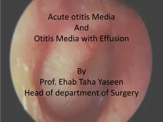
Acute otitis media (AOM) Otitis media with effusion (OME)
Rapid onset of signs and symptoms of inflammation in the middle ear Middle ear effusion (MEE) Bulging or fullness of the tympanic membrane (TM) Erythema of the TM Acute perforation of the TM Otorrhea Otalgia, irritability, and fever
MEE without signs and symptoms of acute inflammation as found in AOM
Proper head and neck examination to identify predisposing conditions 1. Craniofacial anomalies such as those of Down syndrome and Treacher Collins syndrome 2. bifid uvula or a cleft palate 3. Hypernasality indicates velopharyngeal insufficiency 4. Hyponasality may be caused by obstructing adenoids, nasal polyposis or deviated septum
Pneumatic otoscopy; assessment of the TM and its mobility The normal TM is translucent and concave and moves briskly with positive and negative pressure The assessment of the TM should note position, color, degree of translucency, and mobility Reduced or no mobility of TM results from perforation of TM, effusion in the middle ear or increased stiffness due to scarring or increased thickness of the TM
ranges from severely retracted to bulging Severely retracted TM usually is associated with effusion Mild to moderate retraction indicates negative pressure, MEE, or both Fullness and bulging of the TM are caused by increased pressure or fluid, or both
may be caused by thickening or scarring or presence of MEE A red but translucent TM is a typical finding in a crying or sneezing infant A red TM that is full or bulging is a sign of AOM A pink, gray, yellow, or blue retracted TM with reduced or no mobility usually is seen with OME
Immittance Testing (Tympanometry) When otoscopic evaluation is inconclusive or difficult to perform (in children older than 6 months) Screening as well as follow up
Eustachian Tube Function The eustachian tube in the infant is shorter, wider, and more horizontal than in the adult physiologic functions of the eustachian tube pressure Protection Clearance (drainage) pressure regulation regulation (ventilation) (ventilation)
Otitis media was diagnosed in 39% of children by 9 months and 62% of children by 2 years of age. Streptococcus pneumoniae was the single most common bacterial pathogen in AOM Haemophilus influenzae Moraxella catarrhalis
Viral Infection of the upper respiratory tract mucosa initiates the whole cascade of events that finally leads to the development of AOM
Allergy may induce inflammatory swelling of the eustachian tube mucosa Allergies produce inflammatory obstruction of the nose Bacteria-laden allergic nasopharyngeal secretions may be aspirated into the middle ear
1. Age; 6 and 11 months of age, first onset out of this range is a powerful predictor of recurrence 2. Allergy; higher frequency of OME and MEE in allergic children than in nonallergic children 3. Immunocompetence; Children with recurrent otitis media may have a defect in the immune system 4. Cleft Palate/Craniofacial Abnormality; Otitis media is considered universal in infants younger than 2 years of age with unrepaired cleft palate
Environmental risk factors; day care attendance, number of siblings, tobacco smoke exposure, Bottle feeding, birth weight, Socioeconomic status Air pollution
The incidence of AOM is highest during the fall and winter months and lowest during spring and summer months, which parallels the incidence of URI
1. Promotion of breastfeeding 2. Avoidance of supine bottle feeding and pacifier use, 3. elimination of passive tobacco smoke 4. influenza and Streptococcus pneumonia vaccine
Observation Medical treatment Antibiotics Decongestants/Antihistamines Steroids Surgical treatment Myringotomy/Tympanocentesis Myringotomy with Tympanostomy Tube Insertion Adenoidectomy with and without Tonsillectomy
Observation Sever disease 1. moderate to severe otalgia 2. temperatures higher than 39 C orally or 39.5 C rectally, 3. toxic-appearing child
<6 month <6 month 6 6- -24 24 month month > 24 month > 24 month Diagnosis uncertain? Sever disease? Sever disease? Sever disease? Sever disease? Yes Yes No No Yes Yes AB therapy AB therapy Yes Yes No No Observe Observe Diagnosis certain? Diagnosis certain? No No
Amoxicillin is still the first-line antibiotic for non-severe episodes of AOM 90 mg/kg per day in two two divided doses For severe episodes of AOM, Co-amoxiclav for a 10 KG child 5.5 ml of Farmentin 457 q12h If penicillin allergy; cephalosporin Both penicillin and cephalosporin allergy; macrolids
Treatment failure; persistence or recurrence of symptoms and signs 48 to 72 hours after institution of initial treatment Start antibiotic if the patient was under observation Change amoxicillin to co-amoxiclav Change co-amoxiclav to ceftriaxone for 3 days Tympanocentesis should always be considered if the child does not respond to the antibiotic
<6 years old and sever cas10 day course >6years 5-7 day course azithromycin may be given for 1-, 3-, and 5-day courses one dose of IM ceftriaxone may be given neither decongestants or antihistamines, nor their combinations, and steroids are recommended for the treatment of AOM
Antibiotic Prophylaxis 1. Continuous 2. Intermittent Myringotomy or tympanocentesis relief of pain, sample for culture Tympanostomy tube insertion 1. preventive 2. medical treatments failure of recurrent AOM Adenoidectomy may provide modest improvement in children with recurrent AOM but is not recommended as a first-line procedure unless indicated for airway obstruction
Hearing loss level Hearing loss level <20 db 21-39 dB > 40 dB Sever or long standing symptoms (>4 month) hearing loss, language, learning delays, or suspected structural abnormalities of the ear drum Yes No examination at 3- to 6-month intervals surgery
Decongestant/Antihistamine, antibiotics or steroids are not recommended not Autoinflation might be considered for use while awaiting spontaneous resolution of the effusion Tympanostomy tube insertion is the initial preferred procedure Adenoidectomy should not be performed except for a specific indication (i.e., nasal obstruction, chronic adenoiditis) Repeat surgery consists of adenoidectomy plus myringotomy with/without tympanostomy tube insertion
Decongestant/Antihistamine, antibiotics or steroids are not recommended not Autoinflation might be considered for use while awaiting spontaneous resolution of the effusion Tympanostomy tube insertion is the initial preferred procedure Adenoidectomy should not be performed except for a specific indication (i.e., nasal obstruction, chronic adenoiditis) Repeat surgery consists of adenoidectomy plus myringotomy with/without tympanostomy tube insertion
significant erythema ,edema, and tenderness of the postauricular region. This is the typical presentation of acute mastoiditis. Otoscopy revealed findings consistent with acute otitis media.

")


")
")
")