Overview of Radiology: Advancements and Applications
Radiology plays a vital role in modern medicine, enabling visualization of anatomical structures through various imaging techniques. From the accidental discovery of X-rays to the evolution of advanced modalities like CT scans and MRIs, radiology has significantly contributed to diagnostic imaging. Radiologists have specialized in different areas, utilizing specific devices to enhance diagnostic accuracy in fields such as mammography, interventional radiology, and more. Understanding the physical principles of X-ray attenuation is fundamental to interpreting radiological images effectively and making informed medical decisions.

Download Presentation

Please find below an Image/Link to download the presentation.
The content on the website is provided AS IS for your information and personal use only. It may not be sold, licensed, or shared on other websites without obtaining consent from the author.If you encounter any issues during the download, it is possible that the publisher has removed the file from their server.
You are allowed to download the files provided on this website for personal or commercial use, subject to the condition that they are used lawfully. All files are the property of their respective owners.
The content on the website is provided AS IS for your information and personal use only. It may not be sold, licensed, or shared on other websites without obtaining consent from the author.
E N D
Presentation Transcript
Guidelines: Medical radiology Villa Sistemi Medicali SpA Vi l l a Mar ket i ng Di vi si on
Contents Introduction To Radiology Key Components Work Techniques Types Of Exams Types Of Devices Vi l l a Mar ket i ng Di vi si on
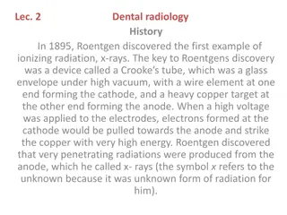
Introduction To Radiology The radiology is used as diagnostic instrument to visualize anatomical structures, that can not be seen from outside and it is a part of a bigger discipline, called DiagnosticImaging The X-rays were discovered accidentally by Wilhelm Conrad R ntgen (German) in 1895 In the course of the century, technologies, means and materials have become refined and have brought the radiology (or better the Diagnostic Imaging) be a particular branch of the medicine A significant evolution of the radiology has been the CT (Computerized Tomography) (Hounsfield et Al, 1972) which still uses collimated X-rays and allows the study of body sections Progressively other diagnostic modalities have been improved, as ultrasound, nuclear medicine, magnetic resonance which use different physical principles from X-rays and allow to obtain different information compared to ones given by radiographic images Vi l l a Mar ket i ng Di vi si on
Introduction To Radiology With the progress of knowledge and of technologies, radiologists have developed a strong specialization in their competences Thanks to X-rays, specialized products were designed, addressed directly to clinical specialist in different fields: Mammography, Interventistic, Angiography, Mineralometry, etc. Each clinical specialty is equipped with devices, which, limiting the application field, increase the diagnostic efficacy of the product Radiological competence can be found also in other specializations as, for example, surgeon, cardiologist, breast specialist, dentists, etc. They use radiological images, but each of them with very specific purpose and needs, that must be understood and supported. Vi l l a Mar ket i ng Di vi si on
Introduction To Radiology Physical principle Radiology is based on the use of X-rays, electromagnetic radiations with wave-length of about 10-10 m X-rays can easily cross each kind of material: they practically go into the object and come out in the opposite side. When X-rays cross an object, they are attenuated by the object itself: the more dense is an object, the higher is its attenuation Air is not very dense: X-rays cross it easily - Water has a middle density: only a part of X-rays can cross it - Lead has a very high density: practically X-rays don t cross it (this is the reason for which the lead is used as protective barrier) - In the human organism there are: elements that are not very dense (soft tissue, muscles, internal organs) and elements very dense (bones) Vi l l a Mar ket i ng Di vi si on
Introduction To Radiology Phy si cal pr i nci pl e X-rays, after having crossed the body, go out and hit a film which is very similar to a normal photographic film The radiographic film is not very sensitive to X-ray , but it s much more sensitive to common light: that s why it is always positioned inside a cassette , which has several purposes: Protect the film from the light Increase the sensitivity of the film, getting it in touch with screens that transform X-rays into visible light - - Increase the mechanical resistance The X-rays are noxious for the organism so it is necessary to limit the exposure of both patients and operators - Obviously the relation risks/benefits is very favorable, in fact the radiological technique, even if is a century old , continues to be used Vi l l a Mar ket i ng Di vi si on
Contents Introduction To Radiology Key Components Work Techniques Types Of Exams Types Of Devices Vi l l a Mar ket i ng Di vi si on
Key Components X-rays are the most popular technology for diagnostic imaging in a wide spectrum of application (bucky tables and remote controlled RF systems) A radiological system is mainly made up of the following elements: Patient positioner - Table - X-ray source - Tube - High voltage Generator - Image receptor - Film, Image Intensifier, DR - In addition, there are: Grid - Collimator - High tension cables - Vi l l a Mar ket i ng Di vi si on
Key Components Table The table is the element which is mainly useful for support and the positioning of the patient Typically the table is also provided with a column, which supports the X-ray tube and allows its positioning as needed Tube Tube Column Column Apollo Moviplan Vi l l a Mar ket i ng Di vi si on
Key Components Table The table is equipped with a device to support and position the cassette: The most simple system is called Cassette Holder and is not used frequently A middle system is called Potter Bucky and it is typically used for bone exams (Moviplan, chest stands) The most sophisticate system is called Spot Film Device and it is typically used for Gastro-Intestinal exams on R/F tables (Apollo, Vision) Potter Bucky Spot Film Device Vi l l a Mar ket i ng Di vi si on
Key Components Tube The X-ray tube is the component which emits the X-rays To emit the X-rays, the tube must be supplied with a voltage from 40 to 150 kV There are several models of tube, in function of the kind of the system, of the application and of workflow The models are different as a function of several parameters: Power Focal spot size Heat storage capacity - - - Vi l l a Mar ket i ng Di vi si on
Key Components Generator The generator generates the high voltage to be applied to the tube to produce X-rays There are several models in function of the power and of the application The rad-only generators are used with tables as with Moviplan for bones applications The R/F generators are used with remote controlled tables (Apollo) and tilting tables (Vision and Viromatic) in gastro-intestinal (GI) and angiographic applications With the console it is possible to set three main parameters: kV (Kilovolt): from 40 to 150 - mA (milliampere): from 10 to 1000 - s (seconds): from 0.001 to 20 Changing those three parameters it is possible to adapt X-ray emission to the organ in exam and to the kind of image that the operator wants to obtain - Vi l l a Mar ket i ng Di vi si on
Key Components Image receptor The radiographic film, similar to a photographic film, is hit by the X-rays and an image with the crossed tissue density can be obtained The Image Intensifier subsystem is used to operate in fluoroscopy mode to convert x-rays into a bright visible light Digital X-ray sensors are used in Digital Radiography, instead of traditional film, and are called Flat Panel Detectors. They can be fixed or portable and contain a layer of scintillator material commonly made by Caesium Iodide (CsI) or Gadolinium oxysulfide (Gadox) Vi l l a Mar ket i ng Di vi si on
Key Components Grid Inside the Potter Bucky or the SFD a component called grid is present and it has the purpose of eliminating the scattered radiation , that is a sort of fog generated by the subject in exam, which affect the image quality The grid is a sort of filter which cleans the image Vi l l a Mar ket i ng Di vi si on
Key Components Collimator The collimator is present on the tube in the point from which X-rays are emitted, and is used to: Limit the size of the irradiated field Project on the patient a luminous square which shows the field size. Reduce the stray radiation in the environment Collimator Vi l l a Mar ket i ng Di vi si on
Key Components HT cables The high tension cables (HT) are used to connect the generator with the tube Their length must be chosen in function of the system composition (14/16 m cables are the most common ones) Vi l l a Mar ket i ng Di vi si on
Contents Introduction To Radiology Key Components Work Techniques Types Of Exams Types Of Devices Vi l l a Mar ket i ng Di vi si on
Work Techniques Parameters meaning kV are representative of single photons energy and therefore of their penetration power. A radiation with high kV can cross denser and/or thicker materials mA are representative of quantity of emitted X-photons and, under the same kV and object in exam, a radiation with higher mA makes the film darker Exposure time (s) can be assimilated to emitted photons quantity: under the same mA, if the exposure time doubles, also the photons quantity doubles Vi l l a Mar ket i ng Di vi si on
Work Techniques Parameters meaning mA and s are therefore comparable in the final effect (film darkening) The mAs (milliampere-second) unit, as result of the product mA*s, is often used For example, if 25 mAs are necessary to obtain a correct exposure, it is possible to use 25 mA for one second, or it is possible to use 250 mA for 0.1 s It is preferable to use high mA and short times to reduce the artifacts due to the movement of the patient or of the organs Vi l l a Mar ket i ng Di vi si on
Work Techniques Three points technique: the operator can set independently kV, mA and s values Two points technique: the operator can set kV and mAs values. The generator sets the highest possible mA value for the kV value selected One point technique: the operator can set only the kV value Zero points technique: automatic setting of kV, mA, s Vi l l a Mar ket i ng Di vi si on
Work Techniques Anatomic programs Allow to set exposure parameters automatically The operator sets the organ to examine, the projection (frontal or lateral) and the patient size The system sets the best kV, mA, sec values in function of the exam Vi l l a Mar ket i ng Di vi si on
Work Techniques Automatic Exposure Control (AEC) Generator accessory Composed of two parts: - AEC interface (it is a board, which is installed into the generator) Measure chamber is placed in the potter bucky or in the SFD immediately above the cassette The X-rays, before hitting the cassette, cross the measure chamber, which emits an electrical signal proportional to the rays quantity which has crossed it - Vi l l a Mar ket i ng Di vi si on
Work Techniques Automatic Exposure Control (AEC) The signal is sent to interface board, which elaborates it and informs the generator to interrupt the exposure when the radiation dose emitted is sufficient to give the correct film darkening Substantially the parameter on which the AEC acts is the exposure time It is called also 1 point technique: the operator can set only the kV value, manually or through an anatomic program (in this case a mixed anatomic technique + AEC is used). The mA value is set automatically with the max possible value for that particular kV value Vi l l a Mar ket i ng Di vi si on
Work Techniques Conventional and HF generators Conventional generator: the wave- shape of the high tension is directly linked to the 50 Hz mains frequency. The kV have a sinusoidal shape which goes from 0 to 100 kV (in example) Conventional X-ray generator kV t X-ray pulses Constant Potential (HF) X-ray generator HF generator: the wave-shape of the high tension is generated by an inverter which works with frequencies of several kHz and it is further levelled so that it is almost constant kV t t Vi l l a Mar ket i ng Di vi si on
Work Techniques Advantages of HF technology Higher efficiency (under the same application, it is sufficient a lower power) Reduction of the patient dose Insensitivity to mains voltage fluctuations Reduction of soft rays , that are the rays generated in the rising and falling portions of the wave. They don t contribute to the image formation, but are noxious for the patient Vi l l a Mar ket i ng Di vi si on
Contents Introduction To Radiology Key Components Work Techniques Types Of Exams Types Of Devices Vi l l a Mar ket i ng Di vi si on
Types Of Exams Radiography: it is comparable to a photo Serial radiography: it is comparable to a sequence of photo taken at very short intervals Fluoroscopy: it is comparable to a movie Pulsed fluoroscopy: it emits X-ray as a series of short pulses instead of a continuous flow, reducing the patient dose Digital radiography or fluoroscopy: it allows to operate in each type of exam in digital way Tomography: it make a sectional image by moving an X-ray source and the film in opposite directions during the X-ray exposure Vi l l a Mar ket i ng Di vi si on
Types Of Exams Radiography It allows to take static images (chest, bones) It puts in evidence fine details It is executed on: - Bucky tables (Moviplan) and chest stands (for chest exams) thanks to the Potter Bucky - Remote controlled tables (Apollo) thanks to under-table SFD - Tilting tables (Vision, Viromatic) thanks to under-table potter bucky Vi l l a Mar ket i ng Di vi si on
Types Of Exams Serial radiography It is used typically for dynamic GI (gastro-intestinal) study More images (from 2 to 6), called divisions, at intervals of 0.5-1 sec. each other, are taken on the same film It is executed on R/F (Radio/Fluoro) tables, tilting or remote controlled, through the SFD Vi l l a Mar ket i ng Di vi si on
Types Of Exams Fluoroscopy It allows to take pictures of dynamic events (gastro- Intestinal) It is not useful to put in evidence details, but to see the evolution of a phenomenon It is executed on R/F (Radio/Fluoro) tables, tilting or remote controlled and on surgical C-arm The image is taken by TV Chain, made up of an II (Image Intensifier) and a CCD camera, and is visualized on the monitor Vi l l a Mar ket i ng Di vi si on
Types Of Exams Digital fluoroscopy Study of dynamic phenomenon/district with middle/high speed (up to 25 img/s) It is used typically for digestive and vascular apparatus study It allows to execute angiographic examination with DSA (Digital Subtraction Angiography) The system is based on a I.I, a high resolution CCD camera and a digital processing system (DIVA-D) Vi l l a Mar ket i ng Di vi si on
Types Of Exams Tomography It is used to take internal organs sections It is based on the principle of cancellation of structures which are outside the slice layer The cancellation is obtained with an defocussing procedure, thanks to a relative movement between the film and the structures which are not interesting For example it is used for kidney, liver and other exams It is often replaced by Computed Tomography and Magnetic Resonance Vi l l a Mar ket i ng Di vi si on
Types Of Exams Tomography X-ray source movement Patient body Focal plane Table surface Cassette movement Film/screen The effect of cancellation of the structures which are outside the focal layer is obtained through the movement in opposite directions of the source and the film respect to the organ. Vi l l a Mar ket i ng Di vi si on
Types Of Exams Tomography It can be executed on: - - Remote controlled tables (standard) Tilting tables, through optional tomographic column - Bucky table through optional tomographic column The simplest systems use a mechanical bar which synchronizes the tube and the potter movement The most sophisticated systems use an electronic system without bar (electronic tomography) Vi l l a Mar ket i ng Di vi si on
Types Of Exams Exposure parameters Radiography, serial Radiography, digital Parameter Fluoroscopy kV 40 120 40 150 mA 10 1000 0,5 10 Times 1 ms 10 sec From few seconds to some minutes Vi l l a Mar ket i ng Di vi si on
Contents Introduction To Radiology Key Components Work Techniques Types Of Exams Types Of Devices Vi l l a Mar ket i ng Di vi si on
Types Of Devices Remote controlled tables Conventional titling tables Bucky tables Chest stands Ceiling tubestands Mobile units Surgical C arms Mammographic units U-arm Digital Radiographic Systems Vi l l a Mar ket i ng Di vi si on
Types Of Devices Remote controlled tables It is the device which allows to execute the widest number of R/F and RAD exams The control console is positioned in a separate room It is typical of Italian and French schools It allows to make the best of digital acquisition systems Vi l l a Mar ket i ng Di vi si on
Types Of Devices Conventional tilting tables It allows to execute R/F exams To execute RAD exams, an under-table potter bucky and a second tube on column or ceiling tubestand are needed The controls are on the table itself It is typical of Anglo-Saxon school Vi l l a Mar ket i ng Di vi si on
Types Of Devices Bucky tables It is a simple device which allows to execute a wide number of exams The tabletop is floating to allows a fast and precise manual positioning It is often coupled with a chest stand to make a general radiography room (general rad) It is often provided with vertical movement thanks to a lift, which allows to lower the tabletop to about 50 cm from floor, to facilitate the access of patients with motion difficulties (elders or with extremities problems) since the device is often used for first study of casualty ward Vi l l a Mar ket i ng Di vi si on
Types Of Devices Chest stands It is an accessory which completes a radiological system (generally based on Moviplan or Vision/Viromatic, sometimes with remote control) It is used to execute chest and spinal column exams with standing patients It can also be used for loaded lower extremities studies Vi l l a Mar ket i ng Di vi si on
Types Of Devices Ceiling tubestands Useful in casualty wards to have 4-side access to the bucky table, without the floor column For lateral projections on remote control tables Vi l l a Mar ket i ng Di vi si on
Types Of Devices Other devices Mobile units: mobile radiographic systems used in trauma and surgery rooms, at patient s bed and also as backup unit in general diagnostic Surgical C arms: radiographic and fluoroscopy systems used in general and vascular surgery, traumatology and orthopedics thanks to their positioning flexibility U-arm Direct Digital Radiographic Systems: systems based on flat panel detectors that can cover virtually all the applications of a modern radiological room assuring flexibility Mammographic units: analog or digital, are available ready for biopsy Vi l l a Mar ket i ng Di vi si on
Types Of Devices Radiological tables and applications Kind RAD R/F Applications Model Villa Bones Gastrointestinal (GI) Angiography Tomography Chest Remote control R/F Apollo Conventional R/F Vision Viromati c Bones Gastrointestinal (GI) Angiography (limited) Tomography Tilting Bones Tomography Bucky Table RAD Moviplan Chest Spinal column Under load inf. extremities Chest stand RAD Chest stand Vi l l a Mar ket i ng Di vi si on
Thank you for your attention! Villa Sistemi Medicali Sales & Marketing Team Vi l l a Mar ket i ng Di vi si on