Parathyroid Hormone (PTH) and Calcitonin in Endocrinology
Explore the role of parathyroid glands, PTH functions, calcitonin, and mineral homeostasis. Learn about calcium regulation, target organs, and hormonal disorders in this comprehensive study of endocrinology.

Download Presentation

Please find below an Image/Link to download the presentation.
The content on the website is provided AS IS for your information and personal use only. It may not be sold, licensed, or shared on other websites without obtaining consent from the author. If you encounter any issues during the download, it is possible that the publisher has removed the file from their server.
You are allowed to download the files provided on this website for personal or commercial use, subject to the condition that they are used lawfully. All files are the property of their respective owners.
The content on the website is provided AS IS for your information and personal use only. It may not be sold, licensed, or shared on other websites without obtaining consent from the author.
E N D
Presentation Transcript
PARATHYROID AND CALCITONIN Zhikal O. Khudhur Zhikal O. Khudhur Endocrinology BIO 414 Spring Semester Week number: 7 Date: / 2024
Objectives Parathyroid glands 1. PTH target organs 2. PTH functions and regulations 3. Calcitonin functions 4. Mineral deposition and resorption 5. Calcium and phosphate homeostasis 6. Calcitriol synthesis 7. Calcitriol functions 8. Parathyroid hormone disorders 9.
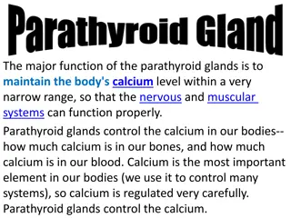
Parathyroid glands The parathyroid glands are partially embedded in the posterior surface of the thyroid gland. There are four parathyroid glands, two on the back of each lobe of the thyroid gland. The hormone they produce is called parathyroid hormone (PTH).
PTH functions and target organs PTH is important for the maintenance of normal blood levels of calcium and phosphate. The target organs of PTH are the bones, small intestine, and kidneys.
PTH regulation and functions PTH secretion is inhibited by hypercalcemia. a high blood calcium level. Secretion of PTH is stimulated by hypocalcemia, a low blood calcium level. PTH raises the blood calcium level by: 1. PTH increases the reabsorption of calcium and phosphate from bones to the blood. 2. The absorption of calcium and phosphate from food in the small intestine, which also requires vitamin D, is increased by PTH. This too raises the blood levels of these minerals. 3. In the kidneys, PTH stimulates the activation of vitamin D and increases the reabsorption of calcium and the excretion of phosphate.
PTH and calcitonin functions Therefore, the overall effect of PTH is to raise the blood calcium level and lower the blood phosphate level. The antagonistic effects of PTH and calcitonin. Together, these hormones maintain blood calcium within a normal range.
Mineral deposition and resorption Mineral deposition (mineralization) is a crystallization process in which calcium and phosphate ions, among others, are taken from the blood plasma and deposited in bone tissue. It begins in fetal ossification and continues throughout life. Mineral resorption is the process of dissolving bone. It releases minerals into the blood and makes them available for other uses. Resorption is carried out by osteoclasts. Hydrogen pumps in the ruffled border of the osteoclast secrete hydrogen ions into the extracellular fluid, and chloride ions follow by electrical attraction. The space between the osteoclast and the bone thus becomes filled with concentrated hydrochloric acid with a pH of about 4. The acid dissolves the bone minerals. The osteoclast also secretes an enzyme called acid phosphatase that digests the collagen of the bone matrix. Acid phosphatase is named for its ability to function in this highly acidic environment.
Calcium and phosphate homeostasis Calcium and phosphate homeostasis depends on a balance between dietary intake, urinary and fecal losses, and exchanges with the osseous tissue. Calcium and phosphate homeostasis is regulated by three hormones: calcitriol, calcitonin, and PTH.
Calcitriol synthesis Calcitriol is a form of vitamin D produced by the sequential action of the skin, liver, and kidneys: 1. Epidermal keratinocytes use ultraviolet radiation from sunlight to convert a steroid, 7- dehydrocholesterol to vitamin D3, and a transport protein carries this to the bloodstream. 2. The liver adds a hydroxyl group to the molecule, converting it to calcidiol [25(OH)D]. 3. The kidney then adds another hydroxyl group, converting calcidiol to calcitriol [1, 25(OH)2D], the most active form of vitamin D.
Calcitriol function The principal function of calcitriol is to raise the blood calcium concentration. It does this by three mechanisms: 1. Increasing calcium and phosphate resorption from the skeleton 2. Promoting the reabsorption of calcium ions by the kidneys, so less calcium lose in the urine. 3. Increasing calcium absorption by the small intestine. (It increases the absorption of phosphate and magnesium ions as well)
Blood calcium homeostasis Thyroid gland releases calcitonin when the blood calcium is high. Parathyroid gland releases PTH when the blood calcium is low.
Parathyroid hormone disorders A. Hypoparathyroidism Hypoparathyroidism is an uncommon condition in which body produces abnormally low levels of PTH It leads to the formation of hypocalcemia B. Hyperparathyroidism Hyperparathyroidism is where the parathyroid glands produce too much PTH. Hyperparathyroidism leads to the formation of hypercalcemia. Types of hyperparathyroidism 1. Primary hyperparathyroidism is caused by parathyroid adenoma or rarely by cancer. The patient will have high PTH, and high calcium level.
Parathyroid hormone disorders 2. Secondary hyperparathyroidism is due to chronic renal failure where the patient has low calcium and high phosphate. Now parathyroid glands persistently produce PTH to maintain calcium level. These patients have high PTH and low or normal calcium 3. Tertiary hyperparathyroidism Tertiary hyperparathyroidism is characterized by excessive secretion of PTH after longstanding secondary hyperparathyroidism, in which hypercalcemia has ensued These patients have high PTH and high calcium levels.
References Greenstein B. & Wood D. F. (2011). The endocrine system at a glance (3rd ed.). Wiley- Blackwell Melmed S. Auchus R. J. Goldfine A. B. Koenig R. Rosen C. J. & Williams R. H. (2020). Williams textbook of endocrinology (14th ed.). Elsevier. Boron W. F. & Boulpaep E. L. (2021). Boron & boulpaep concise medical physiology. Elsevier. Barrett K. E. Barman S. M. Yuan J. X.-J. & Brooks H. (2019). Ganong's review of medical physiology (review questions) (26th ed.). McGraw-Hill Education LLC Molina P. E. (2018). Endocrine physiology (Fifth). McGraw-Hill Education. Zhikal Omar Khudhur 19