Pathogenic Neisseria: Characteristics, Virulence Factors, and Infections
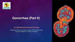
Explore the pathogenic Neisseria species, including characteristics, virulence factors, and infections caused by N. gonorrhoeae. Learn about the general traits, virulence factors, and pathogenicity of Neisseria species, focusing on N. gonorrhoeae with details on its virulence factors and modes of infection transmission. Understand the implications and complications of gonorrhea infections, including common symptoms, affected sites, and potential outcomes. Dive into the world of pathogenic Neisseria and its impact on human health.
Download Presentation

Please find below an Image/Link to download the presentation.
The content on the website is provided AS IS for your information and personal use only. It may not be sold, licensed, or shared on other websites without obtaining consent from the author. If you encounter any issues during the download, it is possible that the publisher has removed the file from their server.
You are allowed to download the files provided on this website for personal or commercial use, subject to the condition that they are used lawfully. All files are the property of their respective owners.
The content on the website is provided AS IS for your information and personal use only. It may not be sold, licensed, or shared on other websites without obtaining consent from the author.
E N D
Presentation Transcript
PHT313 Dr. Hesham Radwan
1. Neisseria species 1. Primary pathogens: a. N. gonorrhoeae (Gonococcus) ALWAYS pathogenic b. N. meningitidis (Meningococcus) May be carried as commensal in nasopharynx flora (10-25% carrier) Grow on chocolate agar at 370C under 5-10% CO2 2. Non-pathogenic (Commensals) Example N. lactamica Grow on ordinary medium at room temperature Habitat of non-pathogenic Upper respiratory, Genitourinary andAlimentary tract 2
Pathogenic Neisseria General characteristics Gram-negative diplococci with adjacent sides flattened (like coffee beans) Non motile Oxidase and Catalase positive Aerobic, capnophilic (5% CO2) and oxidative Requires complex media pre-warmed to 370C Susceptible to cool temperatures, drying and fatty acids Soluble starch added to neutralize fatty acid toxicity Intracellular human-specific pathogen 3
a. Neisseria gonorrhoeae Virulence factors 1. Fimbrae (pili) To adhere to host cells and to each other >100 serotypes known according to pilus protein Has not polysaccharide Capsule 2. Outer membrane proteins (formerly Proteins I, II, & III) Involved in adherence to host cells 3. IgAprotease- cleaves IgAon mucosal surfaces IgAblocks the ability of bacteria to adhere IgAprotease involved in successful colonization 4. Lipopolysaccharide Prevents phagocytosis 4
Pathogenicity Pyogenic infection of columnar and transitional epithelial cells Urethral, endocervix, anal canal, pharynx, conjunctiva 1. Venereal (Sexual Transmitted Disease (STD)) A. Genital infections Gonorrhea B. Extragenital infections Pharyngitis andAnorectal infections 2. Non-venereal Ophthalmia neonatorum Vaginitis in small girls Contaminated toilet seats and contaminated towels 5
Gonorrhea Gonorrhea transmitted by sexual intercourse Gonorrhea is the second most common venereal disease Incubation period: 2 to 7 days Females Males 50% risk of infection after single exposure 20% risk of infection after single exposure Symptomatic infections frequently diagnosed Most initially symptomatic (95% acute) Major reservoir is asymptomatic carriage Major reservoir is asymptomatic carriage Genital infection primary site is cervix (cervicitis), but vagina, urethra, rectum can be colonized Genital infection generally restricted to urethra (urethritis) with purulent discharge and dysuria Ascending infections in 10-20% including salpingitis (Fallopian tubes), tubo-ovarian abscesses, PID, chronic infections can lead to sterility Rare complications may include epididymitis, prostatitis, and periurethral abscesses Disseminated infections more common, including septicemia, infection of skin and joints (1-3%) Bacteremia and Gonococcal arthritis occurs as a result of disseminated gonococcal bacteremia Disseminated infections are very rare Can infect infant at delivery (opthalmia neonatorum) 7
Ophthalmia Neonatorum Contamination of infant s eye during labour through the birth canal of mother In infancy, an eye infection (ophthalmia neonatorum) may occur during vaginal delivery Conjunctivitis with mucopurulent discharge May cause blindness if not treated Infection is preventable with the application of erythromycin eye drops at birth (chlamydia & Neisseria) 8
Laboratory Diagnosis 1. Clinical specimens Gonnorhea Female Cervical discharge in acute and chronic Cervical swab may give positive results Male Acute: Urethral purulent discharge after urinating Chronic: The morning drop or prostatic massage Ophthalmia neonatorum Mucopurulent discharge 2. Direct microscopy (Gram stain) Small, gram-negative diplococci in presence of (WBCs) 9
Laboratory Diagnosis 3. Culture Inoculate specimen on non selective (chocolate agar) or selective media [Thayer-Martin] Incubated at 350C in 5% CO2 The suspected colonies are Gram-negative diplococci with adjacent sides flattened (like coffee beans) 4. Biochemical tests Oxidase + & produce acid from glucose only Fresh growth must be used for testing, because N. gonorrhoeae produces autolytic enzymes 10
Prevention and control Penicillin no longer drug of choice due to: Continuing rise in the MIC Beta-lactamase production (some strains) Ceftriaxone, cefixime or fluoroquinolone combined with doxycycline or azithromycin for dual infections with Chlamydia Chemoprophylaxis against gonorrhea is of little value Chemoprophylaxis against ophthalmia neonatorum with 1% silver nitrate 1% tetracycline or 0.5% erythromycin eye ointment Measures to limit epidemic include education, detection, and follow-up screening of sexual partners, use of condoms No vaccine is available 11
b. Neisseria meningitidis (Meningcoccus) Encapsulated small, gram-negative diplococci Similar to Neisseria gonorrhoeae The main differences are N. gonorrhea Genital tract N. meningitidis Respiratory tract Present None Positive Available Portal of entry Polysaccharide capsule Absent Beta-lactamase Acid from maltose Vaccine Some Negative Not available 12
Pathogenicity: Pili-mediated receptor-specific colonization of cells of nasopharynx Antiphagocytic polysaccharide capsule Allows systemic spread in absence of specific immunity 13 serogroups six of which (A, B, C, W135, X, Y) can cause epidemics Serogroups A, B, C, Y, W135 account for 90% of all infections SerotypesA, B and C are the most common worldwide SerotypeAis common in epidemics inAfrica Toxic effects mediated by hyperproduction of LPS (endotoxin) Other virulence factors as in N. gonnorhea 13
Clinical Infections 1. Meningitis (30-50%) 2. Meningitis with meningococcemia (40%), or 3. Meningococcemia without obvious meningitis (7-10%) Second most common cause of community-acquired meningitis Person-to-person transmission by respiratory droplets Commonly colonize nasopharynx of healthy individuals; highest oral and nasopharyngeal carriage rates in school-age children, young adults and lower socioeconomic groups Requires close contact with infectious person in crowded conditions (e.g. military barracks, prisons, Hajj and other) Requires lack of specific antibody (susceptibility) Incubation period is 2-10 days Symptoms: Fever, headache, stiff neck, nausea, vomiting and purulent meningitis with increased WBCs 15
Laboratory Diagnosis: Specimens CSF appears turbid (meningitis) Blood (meningecoocemia) Nasopharyngeal swabs (carrier) Large numbers (e.g. >107cells/ml) of encapsulated, small, gram-negative diplococci and microscopically in CSF Extracellular and intracellular Culture Transparent, non-pigmented nonhemolytic colonies on chocolate, TM agar with enhanced growth in 5% CO2 Biochemical and Immunologic tests Oxidase-positive Acid production from glucose and maltose Immunologic methods are available for sero-grouping PMN s can be seen 16
Prevention and control 1. Antibiotic prophylaxis is recommended for certain close contacts 2. Avoid crowdedness 3. Vaccination Several vaccines are available to control the disease They are designed to prevent serogroups A, C, Y, and W-135 They are lacking serogroup B antigen 1. Tetravalent Meningococcal Polysaccharide vaccine (MPSV4) Available since 1981 and used for people >55 years 2. Meningococcal Conjugate Vaccine (MCV) When polysaccharides are conjugated to carrier proteins, the polysaccharide antigens become immunogenic in infants and prime for memory anticapsular antibody responses Prophylaxis 17
Treatment Penicillin (drug of choice) Other options: rifampin or sulfonamide (Chemoprophylaxis) 18
Enterobacteriaceae 1. Coliforms (lactose ferementers) Normal inhabitants of GIT of human and animals Source of noscomial infections Opportunistic or cause secondary infections of wounds, urinary and respiratory tracts and the circulatory system e.g. E. coli, Klebsiella E. coli used as biological indicator in water pollution 2. True pathogens (Lactose non-fermenters) Salmonella spp., Shigella spp., Yersinia spp. Certain strains of E. coli (ETEC, EPEC, EIEC, EHEC) 19
Media for isolation Selective differential media for enteric pathogens MacConkey agar EMB agar SS agar Selective by incorporation of dyes and bile salts Differential by incorporation of lactose and/or Fe+3 Fe+3is incorporated to detect H2S Classified as lactose fermenters & non-lactose fermenters 20
I- Escherichia coli General characteristics: Gram-negative Motile rods Non-spore forming,Facultative anaerobic, Oxidase -ve Ferment glucose and lactose Normal flora of intestine Opportunistic pathogens E. coli may be pathogenic inside or outside Intestine Some strains (Pathogenic) cause various forms of gastroenteritis 21
Virulence factors 1. Adhesions (Colonization factors) Pili or fimbriae & nonfimbrial factors 2. Host defense Capsule OMPs are involved in helping the organism to invade by helping in attachment and in initiating endocytosis 3. Exotoxin production (Enterotoxin) Heat-Labile (LT) & Heat-Stable Toxin (ST) (ETEC) Shiga-like toxin (Verotoxin) (EHEC) 4. Endotoxin (Pyrogen) LipidAof LPS causes fever and endotoxic shock 22
Diseases caused by E. coli A. Intestinal: Diarrhea (Toxin and/or adhesion) A. Enterotoxegenic E. coli (ETEC) B. Enteropathogenic E. coli (EPEC) C. Enteroinvasive E. coli (EIEC) D. Enterohemorrhagic E. coli (EHEC) E. Enteroaggregative E. coli (EAEC) B. Extraintestinal: 1. Urinary Tract Infections [UTI] (Pili) 2. Neonatal meningitis (K1 antigen) 3. Sepsis (commonly in debilitated hospitalized patients) Endotoxic shock Due to lipidA(Pyrogen) Fever and sudden hypotension Acquired ingestion of contaminated food and water by 23
Summary of E. coli gastroenteritis Invasion Pathogenesis Site M.O. Diseases Symptoms LT + ST Fluid + electrolyte loss cAMP + cGMP ETEC Small intestine Non invasive Traveler's diarrhea Infant diarrhea Watery cramps, nausea, low grade fever diarrhea, EIEC Invasive Dysentery Large intestine nonfimbrial adhesin (NFA): OMP dysentery-like diarrhea, inflammation, fever Watery diarrhea With mucous without blood or pus With fever & vomiting Hemorrhagic with sever abdominal cramps, diarrhea followed by blood, no fever HUS Watery diarrhea Vomiting Abdominal pain Without inflammation or fever severe EPEC Small intestine NFA: EPEC adherence factor Some reports of shiga-like toxin Cytotoxic shiga-like toxin (verotoxin) intimin Poorly invasive Infantile diarrhea Bloody diarrhoea and haemolytic uraemic syndrome EHEC Large intestine Poorly invasive colitis watery Aute persistent diarrhoea children adults and EAEC Small intestine Production of enterotoxin that similar to ETEC Non invasive in and 24
Urinary Tract Infection (UTI) E. coli is the most common organism causing UTI Community acquired 90% Hospital acquired (50%) UTI is the disease of female (Short urethra) Fecal E. coli acquires pili to colonize mucosa of UT Travel up urethra & infect balder (Cystitis)& sometimes move further up to infect kidney (pyelonephritis) Symptoms:urinary frequency, dysuria, hematuria, pyuria 25
Diagnosis of UTI Specimen MSU (Mid-Stream Urine) Culture On MacConkey agar Gram negative, colonies) Viable count 100,000 (105) cfu/ml urine IMViC + + - - Lactose fermented (Pink 26
Neonatal meningitis E. coli is the second most common cause S. galactaiae (Group B) is the first Occur during the first month of life Lab diagnosis Specimen: CSF Culture: Pink colonies on MacConkey agar (LF) 27
II- Salmonella General characteristics: Gram-negative Enterobacteriaceae Do not ferment lactose and H2S positive Salmonellae live in the intestinal tracts of animals Not considered part of normal intestinal flora in human Always PATHOGENIC to human rods belonging to 28
Classification of Salmonella A. Kaufman-White- Le Minor Classification Based on O and H antigen serotyping 64 O and 114 H variants identified Classified into 9 groups (A-I) according to O antigen Each group can be classified into subgroups according to HAg Salmonella can be detected by its group O antigen and then by its type specific H antigen >2500 known serovars 29
Classification of Salmonella B. US CDC (Center for Disease Control) 1. S.enterica Subdivided into 6 subspecies enterica, salamae, arizonae, diarizonae, indica, houtanae Of these six subspecies, only subspecies enterica is associated with disease in warm-blooded animals S. enterica subsp. enterica serovar Typhi or S. Typhi S. enterica subsp. enterica ser.Typhimurium or S. Typhimurium S. enterica subsp. enterica ser. Enteritidis or S. Enteritidis 2. Salmonella bongori (Subspecies V) 30
Species of Salmonella Two important members of Salmonella causing diseases: A.Salmonella causing enteric fever Salmonella Typhi or Salmonella Paratyphi A,B, and C These organisms penetrate intestinal mucosa Detected in blood, urine and stool B.Salmonella causing food poisoning Salmonella Enteritidis and Salmonella Typhimurium These organisms do not penetrate intestinal mucosa Detected in stoolonly 31
Enteric Fever Typhoid Enteric fevers are severe systemic forms of salmonellosis. Caused by S. typhi whereas a milder form Paratyphoid caused by S. paratyphi A, B or C Mode of Transmission Via fecal-oral route through fecally-contaminated food or water from either chronic carrier or case A few individuals continue to harbour Salmonella in their gall-bladders and intermittently excrete organisms in their faces Temporary excretors: Patients who excrete bacteria for <year Chronic carrier: Patients who excrete bacteria for > year Incubation Period 10-14 day during which bacteria multiplies in Peyer s patches Pass to blood via lymphatics resulting in bacteriaemia in 1stweek In 2ndweek M.O. passes to different organs including peyer s patches causing ulcers, gall bladder, liver, kidney & rarely menings 32
Symptoms of Enteric Fever Symptoms begin after an incubation period of 2 weeks Enteric fevers may be preceded by gastroenteritis, which usually resolves before the onset of systemic disease The symptoms of enteric fevers are nonspecific and include Fever, headache, delirium (sustained fever), malaise and tender abdomen Complications include intestinal hemorrhage and perforation 33
Laboratory diagnosis A. Direct diagnosis: 1. Specimen: Blood during 1st week, urine during 2nd week and stool during 3rd week 2. Isolation of microorganism: I. From blood using blood culture Five to 10 ml of blood is taken during the 1st week of infection, Add to 50-100 ml sterile nutrient broth and incubate at 370C for 24 hrs. Subculture is done on MacConkey's agar which shows colorless colonies in positive case 34
Laboratory diagnosis II. From stool By culture on enrichment medium such as selenite F or tetrathionate broth which inhibits the growth of coliform and allow the growth of Salmonella and Shigella Subculture on MacConkey, SS or DCAagar On MacConkey's agar they give colorless colonies On SS agar they give colorless colonies with black edges due to H2S production Biochemical Reactions: The suspected colonies were subjected to biochemical reactions Oxidase negative, not ferment lactose and sucrose, H2S positive 3. 35
Laboratory diagnosis A. Indirect: Serological diagnosis (Widal Test): In the 2ndweek of the disease, antibodies against Salmonella are present in the patient's serum and can be detected serologically by Widal test (agglutination test) Widal test is positive and valid during 2ndweek Serial dilutions of patient's serum are added to an equal volume of common O and specific H antigens Agglutination of O- antigen and one only of the H-antigens at a titer 1/80 or above is diagnostic 36
Salmonella causing food poisoning The causative agent : S. typhimurium & S. enteritidis Mode of infection: Consumption of contaminated food Food as cakes, pastries and various milk and egg dishes Cattle, sheep, hens, ducks and turkeys are often infected and the organism may contaminate meat and meat products Infective dose 100,000 bacteria Gastric juice is the an important host defense and decreased acidity is a predisposing factor Incubation period is 12-24 hrs and recovery within 4-7 days It is self limiting disease Manifestations include Nausea, vomiting, and abdominal discomfort, non-bloody diarrhea and slight fever 37
I- Shigella General Characteristics Gram negative rods Non motile Non spore-forming Non capsulated Oxidase negative Ferment glucose with acid only Non lactose fermentating organism H2S negative 38
Shigella species Disease: Bacillary dysentery (Shigellosis) S. sonnei is the most common, followed by S. flexneri Mode of Transmission Oral-fecal transmission <200 bacilli are needed for infection in health individuals Incubation periods It varies between 1-3 days Symptoms Ranges from asymptomatic to severe bacillary dysentery Watery diarrhea changing to dysentery with frequent small stools with blood, pus and mucus Fever, tenesmus and abdominal cramps 39
Stages of shigellosis A. Early stage: 1. Ingestion of contaminated food or water 2. Noninvasive colonization and cell multiplication 3. Production of the enterotoxin in the small intestine 4. Watery diarrhea attributed to enterotoxic activity of Shiga toxin (similar to LT of ETEC) 5. Fever attributed to neurotoxic activity of toxin B. Second stage: 1. Adherence to and tissue invasion of large intestine 2. Typical symptoms of dysentery 3. Cytotoxic activity of Shiga toxin increases severity 40
Laboratory identification Specimen: Stool (mucous bloody part of stool) or rectal swap Stool culture Specimen is inoculated in Selenite broth at 370C for 24 h Then subculture on MacConkey or SS On MacConkey agar: they give colorless colonies On SS agar: they give colorless colonies Gram stain Gram negative bacilli, NON MOTILE Biochemical reactions Oxidase negative, ferment glucose, non ferment lactose and H2S NEGATIVE 41
COMMENSAL ENTEROBACTERIAE Opportunistic pathogens and are common cause of nosocomial infections Klebsiella Enterobacter Serratia Proteus Morganella Providencia 1. 2. 3. 4. 5. 6. Lactose Fermnters Non-Lactose Fermnters 42
Klebsiella-Enterobacter-Serratia group General characteristics: These organisms are very similar All are motile except Klebsiella MR negative; VP positive Simmons citrate positive H2S negative Some weakly urease positive Phenylalanine deaminase negative These organisms belonging to Enterobacteriaceae Frequently in large intestine but also found in soil and water Usually opportunistic pathogens Wide variety of infections: primarily pneumonia, wound and UTI 43
Klebsiella pneumoniae Virulence factors 1. Polysaccharide capsule Protects against phagocytosis and antibiotics Makes the colonies moist and mucoid 2. Adhesions Clinical significance Major cause of nosocomial infections Nosocomial Pneumonia Important respiratory tract pathogen outside hospitals 3% of bacterial pneumonia Bloody sputum (50%) (Thick, Jelly and Red Sputum) Septicemia, Meningitis and UTI 44
Proteus-Providencia-Morganella These organisms belonging to Enterobacteriaceae All are normal intestinal flora but also found in soil and water Opportunistic pathogens Non-lactose fermenters Phenylalanine deaminase and urease positive Urease positive after 2-6 hrs (urea NH3+ CO2) All are highly motile Proteus sp. swarming on blood agar Swarming characterized by expanding waves (rings) of organisms over the surface of blood agar 45
Proteus species P. mirabilis and P. vulgaris are widely recognized human pathogens Isolated from urine, wounds and ear and bacteremic infections They found in colon and able to colonize urethra especially in female 1. P. mirabilis causes urinary tract infections (UTI): Proteus sp. produces urease precipitation of calcium and magnesium salts stone which alkalinizes urine formation renal epithelium damage 2. P. vulgaris bacteremia) and UTIs causes nosocomial infections (pneumonia, 46
Laboratory diagnosis Specimen: Urine or Stool. Culture: On MacConkey agar non lactose fermenters colorless colonies On SS agar non lactose fermenters colorless colonies with black center On ordinary media, such as nutrient agar, blood agar, show swarming (successive waves on the surface) due to high motility of Proteus Biochemical reactions: Urease , phenyldeaminase and H2S positive 47
Pseudomonas aeruginosa Gram-negative bacilli belonging to Pseudomonadaceae Motile by single polar flagellum, non spore Non fastidious- Minimal nutritional requirements Produces grape-like odor and blue-green pus and colonies Two types of soluble pigments: 1. Pyoverdin : Yellow-green pigment & fluorescent 2. Pyocyanin: Blue-green pigment and non-fluorescent Strict aerobic, O+/F-, Oxidase and catalase positive Optimum temperature is 370C & able to grow at 420C Resistant to dyes, weak antiseptics and many antibiotics 48
Epidemiology The most important pathogenic is Ps. aeruginosa Opportunistic pathogen Important agent in causing nosocomial infections Ubiquitous in moist environment, hospital, in soil and water Colonized in the intestine in10% of human Represents 10-20% of hospital-acquired infections Transiently colonize respiratory & GIT of hospitalized patients: 1. Treated with broad-spectrum antibiotics 2. Exposed to respiratory therapy equipment 3. Hospitalized for long periods 49
Pathogenesis 1.Adhesins Fimbriae, Pilli, Alginate (mucoid exo-polysaccharide) Alginate slim forms biofilm that protect from antibodies, complement, phagocytosis & antibiotic 2. Invasins a.Proteases - Inactivate IFN and TNF i. Elastase Break down of elastin-containing tissues e.g. blood vessels, lung tissue, skin Cleaves collagen, IgG, IgAand complement) Produce hemorrhagic disseminated infection ii. Alkaline protease (lyses fibrin) b. Hemolysins (phospholipase & lecithinase) & Leukocidin lesions associated with 50


