Pathology Inflammation -1, Acute Inflammation
Acute inflammation is a rapid response to tissue injury, characterized by vascular and cellular changes. Learn about causes, cardinal signs, and nomenclature in this study of acute inflammation.

Download Presentation

Please find below an Image/Link to download the presentation.
The content on the website is provided AS IS for your information and personal use only. It may not be sold, licensed, or shared on other websites without obtaining consent from the author.If you encounter any issues during the download, it is possible that the publisher has removed the file from their server.
You are allowed to download the files provided on this website for personal or commercial use, subject to the condition that they are used lawfully. All files are the property of their respective owners.
The content on the website is provided AS IS for your information and personal use only. It may not be sold, licensed, or shared on other websites without obtaining consent from the author.
E N D
Presentation Transcript
Tishk International University Science Faculty Medical Analysis Department Pathology Inflammation -1, Acute Inflammation Fourth Grade- Spring Semester 2020-2021 Dr. Jalal A. Jalal Assistant Professor of Pathology
Objectives Inflammation Definition Function Causes of acute inflammation Cardinal signs of acute inflammation Vascular changes Cellular changes
Introduction Inflammation: Is the response of vascularized living tissue to injury. Functions to: 1. Dilute toxins 2. Destroy & 3. Isolate offending agent. 4. Initiate repair of tissue
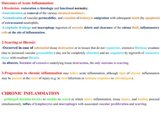
Acute inflammation Immediate and early response to tissue injury. Acute inflammation is of relatively short duration (hours to days) and is primarily characterized by exudation of fluid and plasma proteins, as well as a neutrophilic infiltration.
Causes of Acute inflammation: Causes of Acute inflammation: 1. Infections Infections (bacterial, viral, parasitic) 2. Trauma Trauma 3. Physical and chemical agents Physical and chemical agents (thermal injury, e.g., burns or frostbite; irradiation; chemicals) 4. Tissue necrosis Tissue necrosis (from any cause) 5. Foreign bodies Foreign bodies (splinter, dirt, sutures) 6. Immune reactions Immune reactions (also called hypersensitivity reactions)
Cardinal signs of acute inflammation Cardinal signs of acute inflammation Rubor = redness Tumor = swelling Calor = heat Dolor = pain (described by Celsus 1st. Century AD) Functio laesa = loss of function (added by R. Virchow) Cellulits = acute skin infection commonly caused by Streptococcus pyogenes or Staphylococcus aureus
Nomenclature The inflammation in different tissues employs the tissue name and the suffix -itis e.g. nomenclature used to describe pancreatitis meningitis pericarditis arthritis
Acute inflammation involves: alteration of vascular caliber (vasodilation leads to increased blood flow) changes of microvasculature (increased permeability for plasma proteins and cells) emigration of leukocytes from microcirculation (leukocyte activation leads to elimination of offending agent)
Vascular changes and fluid leakage during acute inflammation lead to Edema in a process called Exudation Edema : excess fluid in interstitial tissue or body cavities, either: 1. Exudate: result of inflammation (increased permeability) high protein and cell debris - content specific gravity >1.020 2. Transudate: result of hydrostatic or osmotic pressure imbalance (ultra filtrate of plasma, no increased vascular permeability) low protein content specific gravity < 1.015 Pus: inflammatory exudate rich in neutrophils, debris of dead cells, microbes
Vasodilation Vasodilation Brief arteriolar vasoconstriction followed by vasodilation Accounts for warmth and redness Opens microvascular beds Increased intravascular pressure causes an early transudate (protein-poor filtrate of plasma) into interstitium permeability still not increased yet) (vascular
Vascular leakage Increased commences Transudate gives way to exudate (protein-rich) Increases interstitial contributing to edema (water and ions) Vascular permeability (leakiness) osmotic pressure
Vascular leakage Several mechanisms known to cause vascular leakage 1. Endothelial cell contraction that widens intercellular gaps of venules (not arterioles, or capillaries) Caused by histamines, bradykinins, leukotrienes which cause an early, brief (15 30 min.) immediate transient response. Most common cause of increased Vascular permeability.
Vascular leakage 2. Severe injuries may cause immediate direct endothelial cell damage (necrosis, detachment) making them leaky until they are repaired (immediate sustained response), or may cause delayed damage as in thermal or UV injury, or some bacterial toxins (delayed prolonged leakage)
Vascular leakage Also marginating and endothelial cell-adherent leukocytes may damage the endothelium through activation and release of toxic oxygen radicals and proteolytic enzymes, making the vessel leaky.
Vascular leakage 3. Increased transcytosis:Certain mediators (VEGF) may cause increased transcytosis via intracellular vesicles which travel from the luminal to basement membrane surface of the endothelial cell. All or any combination of these events may occur in response to a given stimulus
Vascular changes play an important role during acute inflammation Vasodilation, leads to increased blood flow causing redness and warmth (rubor and calor) Increased Permeability, leads to exudation of protein rich fluid into the extravascular space causing swelling (tumor) Concentration of red cells due to fluid loss in small vessels leads to decreased velocity and stasis of the blood flow Leukocyte rolling, adhesion and migration leads to the accumulation of inflammatory cells
Leukocyte cellular events Leukocytes leave the vasculature routinely through the following sequence of events: Margination and rolling Adhesion and transmigration Chemotaxis and activation They are then free to participate in: Phagocytosis and degranulation Leukocyte-induced tissue injury
Margination and Rolling With increased vascular permeability, fluid leaves the vessel causing leukocytes to settle-out of the central flow column and marginate along the endothelial surface Endothelial cells and complementary surface adhesion molecules which briefly stick and release causing the leukocyte to roll along the endothelium until it eventually comes to stop as mutual adhesion reaches a peak leukocytes have
Margination and Rolling Early rolling adhesion mediated by selectin family: E-selectin (endothelium), P-selectin (platelets, endothelium), L-selectin (leukocytes) bind other surface molecules upregulated on endothelium by cytokines (TNF, IL-1) at injury sites (i.e.,CD34,),which are
Adhesion Rolling comes to stop and adhesion results Other sets of adhesion molecules participate: Endothelial: ICAM-1, VCAM-1 (Ig family). Leukocyte: LFA-1, Mac-1, VLA-4 (ICAM-1 binds LFA-1/Mac-1, VCAM-1 binds VLA-4) Ordinarily down-regulated or in an inactive conformation, but inflammation alters this. VLA-4 very late antigen 4 Mac-1 macrophage 1
Transmigration (diapedesis) Transmigration of leukocytes out of the vessel into the interstitial space. Occurs after firm adhesion within the systemic venules and pulmonary capillaries via PECAM 1 (CD31) Must then cross basement membrane, which is degraded by collagenase enzymes.
Transmigration (diapedesis) Early in inflammatory response mostly PMNs, but as cytokine and chemotactic signals change with inflammatory response, endothelial cell expression activates other populations of leukocytes to adhere lymphocytes, etc) progression alteration adhesion of of molecule (monocytes,
Chemotaxis Leukocytes follow chemical gradient to site of injury (chemotaxis) Chemotactic agents: Soluble bacterial products Complement components (C5a) Cytokines (e.g., IL-8) LTB4 (AA metabolite)
Chemotaxis and Activation Leukocytes: extend pseudopods with overlying surface adhesion molecules (integrins) that bind ECM during chemotaxis undergo activation: Prepare AA metabolites from phospholipids Prepare for degranulation and release of lysosomal enzymes (oxidative burst) Regulate leukocyte adhesion molecule affinity as needed.
Summary Inflammation Is the response of vascularized living tissue to injury. According to duration and onset we have acute and chronic inflammation. Cardinal signs of inflammation are Redness, swelling, heat, pain and loss of function. Acute inflammation involves: 1. Vasodilatation. 2. Increased vascular permeability. 3. Emigration and activation of leucocytes.


")
")