Pathology of Gastro-Intestinal Tract GIT
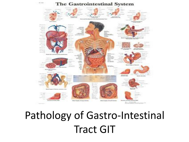
The pathology of the gastrointestinal tract including Fistula, Sinus, Barrett's Esophagus, Gastritis, Acute and Chronic Gastritis, with detailed information on their pathological features. Images provided for visual reference.

Download Presentation

Please find below an Image/Link to download the presentation.
The content on the website is provided AS IS for your information and personal use only. It may not be sold, licensed, or shared on other websites without obtaining consent from the author.If you encounter any issues during the download, it is possible that the publisher has removed the file from their server.
You are allowed to download the files provided on this website for personal or commercial use, subject to the condition that they are used lawfully. All files are the property of their respective owners.
The content on the website is provided AS IS for your information and personal use only. It may not be sold, licensed, or shared on other websites without obtaining consent from the author.
E N D
Presentation Transcript
Pathology of Gastro-Intestinal Tract GIT
Fistula: is an abnormal pathway between two epithelial surfaces.Fistula is an abnormal communication between the lumen of one viscus and the lumen of another viscus or the body surface. Sinus:Sinus is an opening with a blind ending tract, usually lined with granulation tissue.
BARRETTS OESOPHAGUS Pathological features: Columnar metaplasia basal cell hyperplasia Inflammatory changes vary according to the stage of the disease. In early stage, mucosa and submucosa are infiltrated by some polymorphs and eosinophils; in chronic stage, there is lymphocytic infiltration and fibrosis of all the layers of the esophageal wall.
GASTRITIS The term gastritis is commonly employed for any clinical condition with upper abdominal discomfort like indigestion or dyspepsia in which the specific clinical signs and radiological abnormalities are absent.
ACUTE GASTRITIS Acute gastritis refers to a transient inflammation of the gastric mucosa. It is most commonly associated with local irritants such as bacterial endotoxins, caffeine, alcohol, and aspirin. Depending on the severity of the disorder, the mucosal response may vary from moderate edema and hyperemia to hemorrhagic erosion of the gastric mucosa.The complaints of persons with acute gastritis vary from totally unaware of the condition or may report only heartburn or sour stomach to the gastritis that caused by the toxins of infectious organisms, such as the staphylococcal enterotoxins, usually has an abrupt and violent onset, with gastric distress and vomiting ensuing approximately 5 hours after the ingestion of a contaminated food source. Acute gastritis usually is a self-limiting disorder; complete regeneration and healing usually occur within several days.
CHRONIC GASTRITIS: Chronic gastritis is a separate entity from acute gastritis. It is characterized by the absence of grossly visible erosions and the presence of chronic inflammatory changes leading eventually to atrophy of the glandular epithelium of the stomach. The changes may become dysplastic and possibly transform into carcinoma. ETIOPATHOGENESIS. All the causative factors of acute gastritis described above may result in chronic gastritis too. Recurrent attacks of acute gastritis may result in chronic gastritis. Some additional causes are as under: 1. Reflux of duodenal contents into the stomach 2. Associated disease of the stomach and duodenum 3. Chronic hypochromic anemia 4. Immunological factors.
CLASSIFICATION. Based on the type of mucosa affected (i.e. cardiac, body, pyloric, antral or transitional), a clinicopathologic classification has been proposed. 1. Type A gastritis (Autoimmune gastritis). Type A gastritis involves mainly the body-fundic mucosa. It is also called autoimmune gastritis due to the presence of circulating antibodies and is sometimes associated with other autoimmune diseases such as Hashimoto s thyroiditis and Addison s disease. 2. Type B gastritis (H. pylori-related). Type B gastritis mainly involves the region of antral mucosa and is more common. It is also called hypersecretory gastritis due to excessive secretion of acid, commonly due to infection with H. pylori 3. Type AB gastritis (Mixed gastritis, Environmental gastritis, Chronic atrophic gastritis). Type AB gastritis affects the mucosal region of A as well as B types (body-fundic and antral mucosa). This is the most common type of gastritis in all age groups. It is also called environmental gastritis because a number of unidentified environmental factors have been implicated in its etiopathogenesis.
An ulcer: is a discontinuation of the surface epithelium due to necrosis that involving mucosae , muscularis mucosae , the sub-mucosa, or deeper. STRESS ULCERS Stress ulcers are multiple, small mucosal erosions - ulcers, seen most commonly in the stomach but occasionally involving the duodenum.it is of two types: 1.Curling s stress ulcer: refers to gastrointestinal ulcerations that develop in relation to major physiologic stress. Persons at high risk for the development of curling ulcers include those with large surface- area burns, trauma, sepsis, acute respiratory distress syndrome, severe liver failure, major surgical procedures. These lesions occur most often in the fundus of the stomach and proximal duodenum and are thought to result from ischemia, tissue acidosis, and bile salts entering the stomach in critically ill persons with decreased gastrointestinal tract motility.
2. Cushing stress ulcer: consists of gastric, duodenal, and esophageal ulcers arising in persons with intracranial injury, operations, or tumors. They are thought to be caused by hypersecretion of gastric acid resulting from stimulation of vagal nuclei by increased intracranial pressure. These ulcers are associated with a high incidence of perforation. Stress ulcers develop in approximately 5% to 10% of persons admitted to hospital intensive care units. They usually are manifested by painless upper gastrointestinal tract bleeding. Monitoring and maintaining the gastric pH at 3.5 or higher helps to prevent the development of stress ulcers. H2-receptor antagonists, proton pump inhibitors, and mucosal protective agents are used in the prevention and treatment of stress
G/A Acute stress ulcers are multiple (more than three ulcers in 75% of cases). They are more common anywhere in the stomach and the first part of duodenum. They may be oval or circular in shape, usually less than 1 cm in diameter. M/E The stress ulcers are shallow and do not invade the muscular layer. These ulcers commonly heal by complete re-epithelialisation without leaving any scars. Complications: 1. Bleeding 20% 2. Perforation 5% and more in cushing ulcer 3. Obstruction2%
Peptic ulcers: degeneration and necrosis of gastrointestinal mucosa due to exposure to acid-peptic (hydrochloric acid and pepsin) secretions. They can occur at any level of the alimentary tract but they occur most commonly (98-99%) in either the duodenum or the stomach in the ratio of 4:1. Each of the two main types may be acute or chronic. Peptic Ulcers (Gastric Ulcers GU and Duodenal Ulcers DU) Epidemiology: The ratio of DU/ GU = 4/1. Male / female = 3/1 for DU, 2/1 for GU. There is no racial difference in the incidence. It is characterized by remission and relapse.
PATHOGENESIS. i. PU produced by imbalance between gastroduodenal mucosal defenses and the damaging forces especially acid-pepsin secretions ii. Hyperacidity not necessary to be present. In duodenal ulcer there is evidence to support the role of high gastric acid secretion into the stomach at night which takes place under the influence of vagal stimulation, while impaired gastric mucosal defenses (mucous layer) against acid-pepsin secretions seen more in Gastric ulcer with normal or even low acid secretion. iii. H. pylori infection. 80-90 % infected patients not develop peptic ulcers but H. pylori is seen in 95-100% (all) cases of DU and 70% of GU.
The mechanisms by which these bacteria damaging the mucosal defense are include: 1) Gastric mucosal defense is broken by bacterial elaboration of urease, protease, catalase and phospholipase 2) Epithelial injury is also induced by bacterial cytotoxin-associated gene protein (CagA), while vacuolating cytotoxin (VacA) induces release of proinflammatory cytokines such as IL-1, IL-6, IL-8 and tumour necrosis factor- , all of which recruit and activate neutrophils with their damaging properties. 3) H. pylori enhance gastric acid secretion and impair duodenal bicarbonate production. 4) Thrombotic occlusion of surface capillaries is provoked by bacterial platelet activating factor
Other factors may act alone or with H. pylori infection include: 1. NSAIDs-induced mucosal injury. Non-steroidal anti-inflammatory drugs are most commonly used medications in the developed countries and are responsible for direct toxicityby damaging epithelial cells, impairing mucus production, and inhibiting prostaglandin synthesis especially PGE2. 2. hyperacidity (Acid-pepsin secretions). The classic example is zollinger Ellison syndrome in which there are multiple peptic ulcerations in the stomach, duodenum and even jejunum. This is due to excess gastrin secretion by a gastrinomaand excess gastric acid production. Another example is the increase gastric acid secretions in hyperplasia and adenomas of parathyroid glands. 3. Gastritis. Some degree of gastritis is always present in the region of gastric ulcer, though it is not clear whether it is the cause or the effect of ulcer. 4. Other local irritants. Pyloric antrum and lesser curvature of the stomach are the sites most exposed for longer periods to local irritants and thus are the common sites for occurrence of gastric ulcers. Some of the local irritating substances implicated in the etiology of peptic ulcers are heavily spiced foods, alcohol, cigarette smoking, and unbuffered aspirin. 5. Psychological factors. Psychological stress, anxiety, fatigue may exacerbate as well as predispose to peptic ulcer disease. 6. Genetic factors. People with blood group O appear to be more prone to develop peptic ulcers than those with other blood groups. Genetic factor plays greater role in duodenal ulcers as they (DU) run in families, monozygotic twins and association with HLA-B5 antigen. 8. Miscellaneous. Duodenal ulcers have been observed to occur in association with various other conditions such as alcoholic cirrhosis, chronic renal failure, chronic obstructive pulmonary disease COPD, and chronic pancreatitis.
CLINICAL FEATURES. Peptic ulcers are remitting and relapsing lesions. Epigastric pain is the usual clinical presentation for both. The two major forms of chronic peptic ulcers (GU, DU) show variations in clinical features which are as follows: 1. Age. The peak incidence of duodenal ulcer is in 5th decade while that for gastric ulcer is a decade later ie DU occur in younger patients. 2. People at risk. Duodenal ulcer occurs more commonly in people faced with more stress and strain of life (e.g. leaders), while gastric ulcer is seen more often in laboring groups. 3. Periodicity. The attacks in gastric ulcers last from 2-6 weeks, with interval of freedom from 1-6 months. The attacks of duodenal ulcer are classically worsened by work, worry and weather. 4. Pain. In gastric ulcer, epigastric pain occurs immediately or within 2 hours after food and never occurs at night. In duodenal ulcer, pain is more severe, occurs late at night ( hunger pain ) and is usually relieved by food.
5. Vomiting. Vomiting which relieves the pain is a feature in patients with gastric ulcer. Duodenal ulcer patients rarely have vomiting but instead get heart-burn (retrosternal pain) and water brash (burning fluid into the mouth). 6. Haematemesis and melaena. Haematemesis and melaena occur in gastric ulcers in the ratio of 60:40, while in duodenal ulcers in the ratio of 40:60. afraid to eat, while duodenal ulcer patients have very good appetite. 8. Diet. Patients of gastric ulcer commonly get used to a bland diet consisting of milk, eggs etc and avoid taking fried foods, curries and heavily spiced foods. In contrast, duodenal ulcer patients usually take all kinds of diets. 9. Weight. Loss of weight is a common finding in gastric ulcer patients while patients of duodenal ulcer tend to gain weight due to frequent ingestion of food to avoid pain. 10. Deep tenderness. Deep tenderness is demonstrable in both types of peptic ulcers.
Complications: 1. scarring. 2. Bleeding: if the ulcer erodes blood vessels which appear clinically as hematemesis. 3. Iron deficiency anemia due to chronic loss of small amounts of blood. 4. Obstruction: rare, less than 2% of pyloric GU and DU due to edema and scaring 5. Perforation: leading to escape of the gut content into the peritoneal cavity peritonitis presented with acute abdominal pain. 6. Penetration: of the ulcer into the adjacent structures e.g. small intestine. Malignant transformation: not occur with DU but occur in less than 1% of G.U