Pathophysiology of Shock in Cardiovascular Block
Explore the pathophysiological aspects of shock in the cardiovascular block, defining shock and its stages, pathways to decreased tissue perfusion, compensatory mechanisms, and consequences of hypovolemic shock. Learning outcomes include identifying shock categories, understanding tissue perfusion factors, and studying compensatory and irreversible stages. Learning resources from renowned textbooks are provided for further study.

Download Presentation

Please find below an Image/Link to download the presentation.
The content on the website is provided AS IS for your information and personal use only. It may not be sold, licensed, or shared on other websites without obtaining consent from the author. If you encounter any issues during the download, it is possible that the publisher has removed the file from their server.
You are allowed to download the files provided on this website for personal or commercial use, subject to the condition that they are used lawfully. All files are the property of their respective owners.
The content on the website is provided AS IS for your information and personal use only. It may not be sold, licensed, or shared on other websites without obtaining consent from the author.
E N D
Presentation Transcript
Cardiovascular Block Physiology Shock
Intended learning outcomes (ILOs) After reviewing the PowerPoint presentation and the associated learning resources, the student should be able to: Define shock and state the pathophysiological classification of shock. Describe the pathways leading to shock and decreased tissue perfusion. Discuss the stages of a hypovolemic shock. Explain how stage III hypovolemic shock might result in major organs failure. Discuss the different compensatory mechanisms during a hypovolemic shock. Describe the positive feedback mechanisms in the irreversible stage of a hypovolemic shock.
Learning Resources Guyton and Hall, Textbook of Medical Physiology; 12thEdition, Unit IV- Chapters 22 and 24. Linda Costanzo, Physiology, 5thEdition; Chapter 4. Ganong s Review of Medical Physiology; 25th Edition; Section V; Chapter 30.
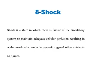
Definition of shock Shock may be defined as a pathophysiological state in which there is widespread, serious reduction of tissue perfusion, which if prolonged, leads to generalized impairment of cellular function. Reduction of tissue perfusion oxygen and nutrients generalized impairment of cellular function failure whole body failure decreased availability of cellular hypoxia and energy deficit cell death organ death.
Pathways leading to decreased tissue perfusion and shock Decreased tissue perfusion can result from hemorrhage / hypovolemic, cardiac failure, or neurologic injury. Decreased tissue perfusion and cellular injury can then result in immune and inflammatory responses. Alternatively, elaboration of microbial products during infection or release of endogenous cellular products from tissue injury cellular activation influences tissue perfusion and the development of shock. endogenous cellular products from tissue injury Endotoxins Activation of receptors
What drives the blood along the blood vessels after it has left the heart?
Systemic hypotension NORMAL BP: 120/80 LOW BP < 90/60 DIASTOLE Arterial pressure = cardiac output x systemic vascular resistance 80 mmHg Diastolic BP 120 mmHg Systolic BP Reduction in either component (cardiac output or vascular resistance) without a compensatory elevation of the other will result in hypotension. SYSTOLE Mean systemic arterial BP ~ 93 mmHg
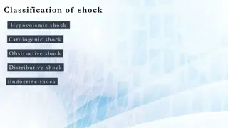
Causes and classification of shock CAUSES SYMPTOMS AND SIGNS TYPE Bleeding (internal/external), dehydration (sever voming, sever diarrhea), plasma loss (as in burns) volume decreased cardiac output hypotension; weak but rapid pulse; cool, clammy skin; rapid, shallow breathing; anxiety, altered mental state Hypovolemic shock low blood hypotension Heart problems (e.g., myocardial infarction, heart failure; cardiac dysrhythmias) decrease in stroke volume decreased cardiac output hypotension decreased contractility as for hypovolaemic shock + distended jugular veins & may be absent pulse Cardiogenic shock Circulatory obstruction (e.g., constrictive pericarditis, cardiac tamponade, tension pneumothorax, pulmonary embolism reduced blood flow to lungs cardiac output hypotension as for hypovolaemic shock + distended jugular veins & pulsus paradoxus (in cardiac tamponade). Obstructive shock decreased Septic shock: infection activation of NOS in macrophages vasodilation hypotension Anaphylatic shock: allergy (release of histamine vasodilation decreased hypotension Neurogenic shock: spinal injury and motor reflexes vasomotor tone vasodilation peripheral vascular resistance release of bacterial toxins production of NO decreased vascular resistance Septic shock: hypotension; fever; warm, sweaty skin Distributive shock Anaphylatic shock: skin eruptions; breathlessness, coughing; localized edema; weak, rapid pulse Vasogenic, resistance vascular Low- resistance shock loss of autonomic reduction of decrease in hypotension Neurogenic shock: as for hypovolemic except warm, dry skin peripheral
Stages of hypovolemic shock STAGE I COMPENSATORY STAGE 10-15% blood loss In healthy individuals, acute blood loss of 10 15 % of the normal blood volume (e.g., blood donation) results in activation of the sympathetic nervous system. Compensation is achieved acutely by: Increase in heart rate. Constriction of the arterioles. Thus, cardiac output is normal or slightly reduced especially when the patient assumes the erect position. Symptoms and signs: Arterial pressure is maintained; pressure decline when the patient assumes the erect position Increase in heart rate Paleness Anxiety See opposite figure.
Stages of hypovolemic shock STAGE II; PROGRESSIVE STAGE (maximal mobilization of compensatory mechanisms) 20 40% blood loss When 20-40% of blood volume are lost, cardiac output cannot be maintained and falls markedly, even in the recumbent position. Accordingly, there will be massive activation of the sympathetic nervous system. This results in: intense arteriolar constriction in the vascular beds of the kidney, gut, and skin redistribution of blood flow (i.e., centralization of blood flow) with larger fractions going to the vital organs (heart and brain) and smaller fractions going to the kidney, gut, and skin. Despite intense arteriolar constriction, systolic arterial BP declines by 10 to 20 mm Hg. generalized venoconstriction (increasing blood volume in the central circulation and tending to sustain venous return) Fluid also moves from the interstitial to the intravascular compartment (i.e., reabsorptionof tissue fluids).
Stages of hypovolemic shock STAGE II PROGRESSIVE STAGE, continued Symptoms, signs, and complications Systolic arterial blood pressure declines by 10- 20 mm Hg Tachycardia (heart rate increases and pulse is thready and weak) Skin is pale and cool (peripheral pallor is common) Deterioration of the mental state because the brain is getting less oxygen. Patient looks weak, tired and drowsy. Patient may show anxiety, aggressiveness, and restlessness. Thirst. Oliguria (urine output is decreased). Angina may occur in patients who have intrinsic coronary vascular disease. pH drops and metabolic acidosis develops due to increased anaerobic glycolysis.
STAGE III; REFRACTORY STAGE Irreversible stage Decompensated stage Stages of hypovolemic shock > 40% blood loss The compensatory mechanisms are maximally mobilized in stage II. Thus, small additional losses of blood (>40 %) can result in life-threatening reductions in cardiac output, blood pressure, and tissue perfusion. Blood flow to heart, brain and kidney is further reduced severe ischemia and irreversible tissue damage in impaired organ function and death. The severe vasoconstriction may itself become a complicating factor and initiate a vicious cycle (see later). this may result Thus, stage III is complicated by major organs failure.
Stages of hypovolemic shock STAGE III REFRACTORY STAGE, continued > 40% blood loss The factors that determine the ultimate outcome include: Duration of stage III. Severity of tissue anoxia. Age and condition of the patient.
Cardiovascular. Respiratory. Endocrine. Renal. Others.
Reduction in arterial MAP during hemorrhage decreases the stimulation of the baroreceptors in the carotid sinuses and aortic arch Sympathetic Parasympathetic (vagal) activity activity and activation of adrenal medulla MAP HR SV TPR Flow to the heart and brain is maintained; reductions in flow to other organs.
Reductions in mean arterial blood pressure (MAP) below 60 mmHg do not evoke any additional responses through the baroreceptor reflex. However, low MAP indirectly stimulates peripheral chemoreceptors (aortic bodies, carotid bodies, heart) that sense changes in pO2, pCO2, and pH through tissue hypoxia and lactacidosis. This results in: Enhancement of the existing tachycardia and vasoconstriction Respiratory stimulation tachypnea
Vasoconstriction (arteriolar constriction): this increases TPR and hence MAP. It is produced by: Baroreceptor reflex. Chemoreceptor reflex. Noreadrenaline-adrenaline vasoconstrictor mechanism (due to activation of adrenal medulla). Vasoconstriction is also produced by vasopressin- vasoconstrictor mechanism (in subsequent slides). Vasoconstriction is marked in: Skin: cold, pale. kidneys: drop in GFR & urine volume. Viscera. Flow to the heart and brain is maintained; reductions in flow to other organs.
Venoconstriction: It is produced by: Baroreceptor reflex. Chemoreceptor reflex. Noreadrenaline-adrenaline vasoconstrictor mechanism (due to activation of adrenal medulla). Venoconstriction is also produced by vasopressin. Venoconstriction is important in: Shifting blood from the venous reservoir into the circulation. Maintaining filling pressure of the heart.
Tachycardia: produced by: Baroreceptor reflex. Chemoreceptor reflex. Increased sympathetic activity. Tachypnea: caused by activation of chemoreceptor reflex and sympathetic over activity. Importance: Increase O2delivery. Increase thoracic pump activity help venous return. The increased sympathetic activity also causes restlessness skeletal muscle activity help venous return.
Control Haemorrhage Reflex Compensation Heart rate Stroke volume EDV Cardiac output Total peripheral resistance Mean arterial blood pressure CO
Control Haemorrhage Reflex Compensation Mean arterial blood pressure Brain Resistance Gut Brain Blood Flow Gut This can Become problematic. Flow to the heart and brain is maintained; reductions in flow to other organs.
1. Release of hormones that initiate vasoconstriction. 2. Renal conservation of salt and water. 3. Stimulation of hematopoeisis by erythropoietin.
Endocrine and renal compensatory mechanisms Stimulation of sympathetic nervous system norepinephrine from the adrenal medulla (sympatho-adrenal axis) . Epinephrin is released almost exclusively from adrenal medulla where as norepinephrin is released from adrenal medulla and sympathetic nerve endings. Vasoconstriction. release of epinephrine, ADH is actively secreted by the posterior pituitary gland in response to hemorrhage. Thus, hemorrhage stimulates posterior pituitary release of vasopressin (ADH). ADH is potent vasoconstrictor. It also stimulates reabsorption of water. Circulating vasoconstrictors synthesis and renal perfusion apparatus generates angiotensin II Angiotensin II is a powerful vasoconstrictor secretion of renin from juxtaglomerular
Endocrine and renal compensatory mechanisms secretion of glucocorticoids by adrenal cortex This helps to maintain blood glucose Stimulation of hematopoiesis blood volume & hypoxia synthesis of erythropoeitin (EPO) hematopoiesis.
Endocrine and renal compensatory mechanisms Vasopressin blood volume Angiotensin II salt reabsorption arterial pressure. arterial pressure in kidney glomerular filtration rates and thereby decreases the excretion of water and electrolytes directly water reabsorption arterial pressure. release of aldosterone blood volume Renal conservation of salt and water decreases
Responses activated within hours: Increased movement of interstitial fluid into capillaries (capillary fluid shift). Increased 2,3 DPG concentration in RBCs: This is important to help Hb deliver more O2to the tissues (shift O2dissociation curve to the right). Responses activated within hours-days: Restoration of circulatory plasma volume. This takes 12-72 hrs after moderate hemorrhage. Restoration of plasma proteins: this occurs in 2 stages: Rapid entry of preformed albumin from extracellular stores. Hepatic synthesis of proteins over 3-4 days. Restoration of RBCs: this occurs in 2 stages: Increase RBCs count in response to erythropoietin within 10 days. Restoration of red cell mass within 4-8 weeks.
In the irreversible stage of hypovolemic shock, a series of positive feed back mechanisms take place leading to further deterioration in cardiac output and MAP and more tissue hypoxia. This depends on the amount of blood lost. When blood loss is excessive and not immediately replaced, and proper treatment is delayed, this stage is reached and the patient dies. In this stage, there is also failure of compensatory mechanisms.
The severe vasoconstriction may itself become a complicating factor and initiate a vicious cycle. Impaired coronary perfusion depression of cardiac function output. Reduced blood flow to the vasomoter centers in the medulla depresses the activity of compensatory reflexes. Prolonged renal ischemia acute tubular necrosis lead to prolonged post-shock renal insufficiency. Bowel ischemia breakdown of the mucosal barrier. This may lead to entry of bacteria and toxins into the circulation. cardiac ischemia further lowering of cardiac may
In the irreversible stage of hypovolemic shock, a number of positive feedback mechanisms lead to further drop in cardiac output and MAP. Impaired coronary perfusion function further lowering of cardiac output. Reduced blood flow to the vasomoter centers in the medulla failure depression of the activity of compensatory reflexes cardiac function further lowering of cardiac output. Bowel ischemia breakdown of the mucosal barrier. This may lead to entry of bacteria and toxins into the circulation further lowering of cardiac output. Generalized cellular deterioration: Mitochondrial activity inside the cells in ATP. of cellular metabolism, especially glucose in ATP. in active transport of Na+and K+across the cell Na+accumulation inside the cell. Rupture of many lysosomes. cardiac ischemia depression of cardiac vasomotor depression of depression of cardiac function
Intended learning outcomes (ILOs) After reviewing the PowerPoint presentation and the associated learning resources, the student should be able to: Define shock and state the pathophysiological classification of shock. Describe the pathways leading to shock and decreased tissue perfusion. Discuss the stages of a hypovolemic shock. Explain how stage III hypovolemic shock might result in major organs failure. Discuss the different compensatory mechanisms during a hypovolemic shock. Describe the positive feedback mechanisms in the irreversible stage of a hypovolemic shock.

")


 below 60")
: this")


")