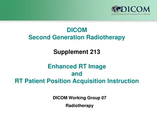
Patient Radiation Dose Reporting Supplement
Patient Radiation Dose Reporting Supplement (P-RDSR) is a DICOM supplement developed by Working Group 28 (WG-28-Physics) on May 31, 2016. It focuses on storing and exchanging patient organ dose calculations for various medical imaging procedures. The supplement aims to enhance the utilization of data related to radiation dose estimation, including radiation beam characteristics, patient/organ models, and dose calculation methods. The goal is to provide a comprehensive framework for sharing dose information across different modalities and calculation scenarios.

Download Presentation

Please find below an Image/Link to download the presentation.
The content on the website is provided AS IS for your information and personal use only. It may not be sold, licensed, or shared on other websites without obtaining consent from the author.If you encounter any issues during the download, it is possible that the publisher has removed the file from their server.
You are allowed to download the files provided on this website for personal or commercial use, subject to the condition that they are used lawfully. All files are the property of their respective owners.
The content on the website is provided AS IS for your information and personal use only. It may not be sold, licensed, or shared on other websites without obtaining consent from the author.
E N D
Presentation Transcript
Supplement 191: Patient Radiation Dose Reporting (P-RDSR) Supplement is developed by DICOM Working Group 28 (WG-28-Physics) 31 May 2016
Patient Radiation Dose SR (P-RDSR) Current Radiation Dose SR contains only information about the x-ray system or information the x-ray system can determine, e.g.: radiation output, geometry, x-ray source, detector system, etc. Estimation of patient/organ dose requires knowledge of: Radiation beam characteristics that interact with patient Models of the patient/organs Models of radiation interaction within the patient Methods to do patient dose estimations are being developed and improved continuously storage of these estimations in a different object would allow more versatile utilization of the data DICOM WG-28 / Supp 191 2 31 May 2016
Patient Radiation Dose SR (P-RDSR) Goals of the Patient Dose SR Store the results of Patient Organ Dose calculations: of a SINGLE procedure or MULTIPLE procedures including one or more modalities and procedure steps/phases of one or more organs by one or more calculation methods Exchange intermediate results with peers Examples of modalities and procedures: Single modality, single procedure step CT internal organ dose (Stochastic) eye dose (Deterministic) XA/RF (one time point) skin dose map (Deterministic) internal organ dose (Stochastic) Mammography glandular dose (Stochastic) Projection x-ray (CR/DX) entrance skin dose (Deterministic) internal organ dose (Stochastic) NM/PET internal organ dose (Stochastic) Multi-modality, single procedure step SPECT and PET/CT internal organ dose (Stochastic) Single modality, multi-procedure step CT internal organ dose (Stochastic) eye dose (Deterministic) XA/RF (multi time points being combined) skin dose map (Deterministic) XA localization / disgnostic plus intervention skin dose map (Deterministic) internal organ dose (Stochastic) Mammography/Projection X-ray Tomosynthesis DICOM WG-28 / Supp 191 3 3 31 May 2016
Patient Radiation Dose SR (P-RDSR) Data flow simple case Calculation Method 1 Radiation Dose SR 1 Patient Dose SR 1 radiation radiation Organ 1 Dose Estimate Group #1: Organ 1 RDSR Source 1 Method 1 DICOM WG-28 / Supp 191 4 31 May 2016
Patient Radiation Dose SR (P-RDSR) One RDSR, multiple organs Calculation Method 1 Radiation Dose SR 1 Patient Dose SR 1 radiation radiation Organ 1 Dose Estimate Group #1: Organs 1, 2, 3 RDSR Source 1 Method 1 Organ 2 Organ 3 DICOM WG-28 / Supp 191 5 31 May 2016
Patient Radiation Dose SR (P-RDSR) Multiple RDSRs, multiple organs Calculation Method 1 Radiation Dose SR 1 Patient Dose SR 1 radiation radiation Organ 1 Dose Estimate Group #1: Organs 1, 2, 3 RDSR Source 1 Method 1 Dose Estimate Group #2: Organs 1, 2, 3 RDSR Source 2 Method 1 Radiation Dose SR 2 Organ 2 radiation Or combining RDSRs: Dose Estimate Group #1: Organs 1, 2, 3 RDSR Sources 1, 2 Method 1 Organ 3 DICOM WG-28 / Supp 191 6 31 May 2016
Patient Radiation Dose SR (P-RDSR) Multiple RDSRs, multiple organs, multiple methods Calculation Method 1 Radiation Dose SR 1 Patient Dose SR 1 radiation radiation Organ 1 Dose Estimate Group #1: Organs 1, 2, 3 RDSR Source 1 Method 1 Dose Estimate Group #2: Organs 1, 2, 3 RDSR Source 2 Method 1 Dose Estimate Group #3: Organs 1, 2, 3 RDSR Source 1 Method 2 Dose Estimate Group #4: Organs 1, 2, 3 RDSR Source 2 Method 2 Calculation Method 2 Radiation Dose SR 2 Organ 2 radiation Organ 3 Or combining RDSRs: Dose Estimate Group #1: Organs 1, 2, 3 RDSR Sources 1, 2 Method 1 Dose Estimate Group #2: Organs 1, 2, 3 RDSR Sources 1, 2 Method 2 DICOM WG-28 / Supp 191 7 31 May 2016
Patient Dose Determination: Data Flow Requirements Modality Frame of Reference (FOR) Modality Image radiation Radiation Dose SR Radiation Exposure Information - X-Ray exposure techniques - Table, Gantry Angle, Beam Geometry, collimation - Dose measure: CTDI, DAP, ... X-Ray Equipment Organ Dose Reporter System Signifies part of Supplement 191 Patient RDSR DICOM WG-28 / Supp 191 8 31 May 2016
Patient Dose Determination: Data Flow Requirements Modality Frame of Reference (FOR) Modality Image radiation Radiation Dose SR Radiation Exposure Information - X-Ray exposure techniques - Table, Gantry Angle, Beam Geometry, collimation - Dose measure: CTDI, DAP, ... X-Ray Equipment Organ Dose Reporter System Equipment Information - Table dimension - Attenuating material, Patient Model Patient Model FOR Registration IOD Patient Registration Information - Patient position on table - Fiducials - Registration matrix between Patient FOR and Equipment FOR Signifies part of Supplement 191 Patient RDSR DICOM WG-28 / Supp 191 9 31 May 2016
Patient Dose Determination: Data Flow Requirements Modality Frame of Reference (FOR) Modality Image radiation Radiation Dose SR Radiation Exposure Information - X-Ray exposure techniques - Table, Gantry Angle, Beam Geometry, collimation - Dose measure: CTDI, DAP, ... X-Ray Equipment Patient Dose Surface 3D map/view Organ Dose Reporter System Equipment Information - Table dimension - Attenuating material, Patient Model Patient Model FOR Patient Dose 2D view/map (e.g. iso- dose map) Patient Dose SR radiation Registration IOD Patient Registration Information - Patient position on table - Fiducials - Registration matrix between Patient FOR and Equipment FOR Calculated data for documentation and reference to images and RDSR s Signifies part of Supplement 191 Patient RDSR DICOM WG-28 / Supp 191 10 31 May 2016
Registration between Patient Model and RDSR Case #1 Patient Images FOR X-Ray Images radiation A B M M M T x x Radiation Dose SR (RDSR) X-Ray Equipment 11 12 13 x A B M M M T - - - Table, position Gantry Angle Beam Geometry, collimation y y 21 22 23 y = Registration A B M M M T z z 31 32 33 z 0 0 0 1 1 1 Registration matrix between: - each RDSR FOR and - the Patient Model FOR Patient Model FOR Patient Model (3D image) DICOM WG-28 / Supp 191 11 31 May 2016
Registration between Patient Model and RDSR Case #2 FOR TABLE TOP HEAD Equipment Fiducials radiation A B M M M T x x Radiation Dose SR (RDSR) X-Ray Equipment 11 12 13 x A B M M M T - - - Table position vs. Isocenter Gantry Angle vs. Isocenter Beam Geometry, collimation y y 21 22 23 y = Registration A B M M M T z z 31 32 33 z 0 0 0 1 1 1 Registration matrix between: - each RDSR Fiducial FOR and - the Patient Model FOR Patient Model FOR Patient Model (3D image) DICOM WG-28 / Supp 191 12 31 May 2016
Registration between Patient Model and RDSR Case #3 FOR TABLE TOP HEAD Equipment Fiducials radiation A B M M M T x x Radiation Dose SR (RDSR) X-Ray Equipment 11 12 13 x A B M M M T - - - Table position vs. Isocenter Gantry Angle vs. Isocenter Beam Geometry, collimation y y 21 22 23 y = Registration A B M M M T z z 31 32 33 z 0 0 0 1 1 1 FOR Registration matrix between: - each RDSR Fiducial FOR and - the Patient Fiducial FOR PATIENT TOP HEAD Patient Fiducials Note: the patient fiducials can be defined through a manual measurent on the actual patient landmarks, or through image- based measurements of landmarks visible on the X-Ray images. DICOM WG-28 / Supp 191 13 31 May 2016
Registration between Patient Model and RDSR Case #4 No X-Ray Images No Equipment Fiducials radiation Radiation Dose SR (RDSR) X-Ray Equipment - - - Table position vs. Isocenter Gantry Angle vs. Isocenter Beam Geometry, collimation Free Text Comment: E.g. Distance from Patient Head to Table Top Head is 15 cm Registration No Patient Model DICOM WG-28 / Supp 191 14 31 May 2016
Patient Radiation Dose SR (P-RDSR) Patient Dose SR IOD Templates TID 1002 Observer Context TID prdsrT01 Patient Radiation Dose TID 1204 Language of Content Item and Descendants TID prdsrT02 Dose Estimate Group TID prdsrT04 Dose Estimate Methodology TID prdsrT05 Dose Estimate Parameters TID prdsrT03 Dose Estimate Representation DICOM WG-28 / Supp 191 15 31 May 2016
Patient Radiation Dose SR: Detailed Structured Content radiation Ref. 1-N Source Radiation Dose SR TID prdsrT01 Patient Radiation Dose 1 Patient Dose Model 1 - Model Type - Radiation Transport Type - Model Reference TID 1204 Language of Content Item and Descendants Ref. 1 1 Patient Model Data Patient Radiation Dose Report 1 - Group Name - Source Category 1 1 Patient Model Demographics 1 Model Age, sex, weight, height TID prdsrT04 Dose Estimate Methodology 1 TID 1002 Observer Context 1 1-N 1 Registration with RDSR Registration Method 1 Ref. Fiducials Ref. 1 Registration TID ptdsrT02 Dose Estimate Group Dose Estimate Methodology 1-N 1 - Material Category - Equivalent Material - Equivalent Thickness - Description X-Ray Beam Attenuator Information 1 1-N 1 1 - Radiation Transport Type - Model Reference Attenuator Model Dose Estimate Group 1 - Registration Method - Fiducials - Registration Ref. Ref. 1 1 Registration with RDSR 1-N 1 Radiation Dose Estimate Method Radiation Dose Estimate Method Type TID prdsrT05 Dose Estimate Parameters Radiation Dose Estimation Parameter Dose Estimate Parameters 1 1-N 1 Legend: TID prdsrT03 Dose Estimate Representation 1 Dose Estimate Representation 1 1-N Template Distribution Model Ref. 1 Container Instance - Organ Name - Absorbed Dose - Equivalent Dose - Uncertainty info 1-N Organ Content Items 1-N 1 Organ Dose Information DICOM WG-28 / Supp 191 16 31 May 2016
Author Contacts Donald Peck, donaldp@rad.hfh.edu (editor) Annalisa Trianni, trianni.annalisa@aoud.sanita.fvg.it Heinz Blendinger, heinz.blendinger@siemens.com Francisco Sureda, francisco.sureda@med.ge.com Steve Massey, steve.massey@pacshealth.com Ioannis Sechopoulos, ioannis.sechopoulos@radboudumc.nl Ting Lu, ting.lu@bayer.com DICOM Secretariat: dicom@medicalimaging.org Thank you for your attention! DICOM WG-28 / Supp 191 17 31 May 2016

")
")
")
")
")
")


")