Pediatric Shock: Types, Characteristics, and Therapeutic Considerations
Explore the types and characteristics of pediatric shock, review therapeutic considerations for different kinds of shock, and learn about the nuances of documenting pediatric shock cases. The content covers definitions, contributors to shock, compensatory responses, stages of shock, and various types of shock such as septic, hypovolemic, hemorrhagic, cardiogenic, distributive, and obstructive shock.

Download Presentation

Please find below an Image/Link to download the presentation.
The content on the website is provided AS IS for your information and personal use only. It may not be sold, licensed, or shared on other websites without obtaining consent from the author. If you encounter any issues during the download, it is possible that the publisher has removed the file from their server.
You are allowed to download the files provided on this website for personal or commercial use, subject to the condition that they are used lawfully. All files are the property of their respective owners.
The content on the website is provided AS IS for your information and personal use only. It may not be sold, licensed, or shared on other websites without obtaining consent from the author.
E N D
Presentation Transcript
Pediatric Shock Theodore K. M. DeMartini, MD Dec 7, 2023
Objectives Describe types and characteristics of pediatric shock Briefly review therapeutic considerations for different kinds of shock Discuss nuances of documentation of pediatric shock
Shock: definition SHOCK HYPOTENSION
Shock: definition HYPOTENSION SHOCK
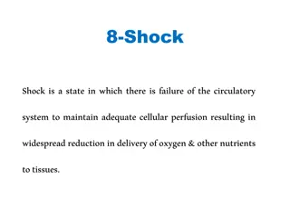
Shock: definition Inadequate oxygen delivery to meet demand of tissues on a macro and a micro level DO2 < VO2 DO2 = CO x CaO2 DO2 = (HR x SV) x [(SaO2 x Hgb x 1.36)+(PaO2 x 0.003)] BP = CO x SVR
Shock: contributors Heart Rate: Bradycardia: high vagal tone, heart block Tachycardia: SVT, V tach, JET Stroke Volume Decreased Preload: hypovolemia, AV desynchrony, impaired venous return Decreased Contractility: myopathies, ischemia, medication effects Increased Afterload: generally relative to contractility
Shock: contributors SaO2/PaO2: 5 causes of hypoxemia Hemoglobin concentration: phlebotomy, hemorrhage, marrow failure Failure of normal blood distribution
Compensatory Responses Effective blood volume Vasoconstriction Decreased renal losses Fluid re-distribution to intravascular space Cardiac Performance Increase HR Increase Contractility Increase Preload Vital Organ Perfusion Oxygen offloading
Stages of Shock Compensated No hypotension Uncompensated Irreversible
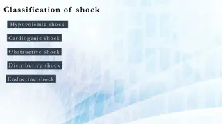
Types of Shock Septic Hypovolemic Hemorrhagic Cardiogenic Distributive Obstructive Type Preload Afterload Contractility Hypovolemic Cardiogenic Distributive , , or Obstructive
Septic Shock Causes: sepsis Mixture of several shock types: Cardiogenic: acute systolic insufficiency Distributive: vasoplegia Hypovolemic: increased capillary permeability leads to fluid shifting to extravascular space
Septic Shock Presentation: variable Cold: poor perfusion, low-normal blood pressure Low CO, high SVR Warm: normal to overly perfused, flashy, low BP (esp diastolic) Normal to low CO, low SVR
Septic Shock: diagnosis SIRS criteria SIRS + suspected/confirmed infection = sepsis Severe sepsis: signs of end-organ perfusion impairment Septic shock: requiring vasoactive infusions to support perfusion qSOFA (adults) Positive if 2/3 variables present: GCS <15, RR >21, SBP <101 Less sensitive than SIRS NOT recommended by Surviving Sepsis guidelines 2021
Septic Shock: pathophysiology Infections trigger innate immune system via pathogen- associated molecular patterns (PAMPs) or damage- associated molecular patterns (DAMPs) The innate immune system then releases pro-inflammatory cytokines (TNF- , IL-1, IL-6) to activate leukocytes, the complement system, and endothelial adhesion molecules Sepsis is an exaggerated response of the above mechanisms
Septic Shock: pathophysiology Inflammation due to sepsis activates the coagulation cascade Can vary from mild thrombocytopenia to disseminated intravascular coagulation Microthrombi form in tissues Fibrinolysis is inhibited Activated Protein C is downregulated, leading to ongoing immune over-activity
Septic Shock: pathophysiology After initial immune hyperactivity, prolonged state of immunosuppression occurs T cell apoptosis and decreased response to cytokines Neutrophils have fewer cytokine receptors
Septic Shock: pathophysiology Decreased delivery of O2: Cardiomyopathy due to cytokines (TNF- , IL-1 ), decreased cardiac myocyte function and mitochondrial function. Systolic and diastolic dysfunction is seen Arterial and venous dilation: decreased BP and venous return/preload Capillary leak due to loss of endothelial barriers Microthrombi preventing delivery to certain tissues Impaired O2utilization: Mitochondrial dysfunction due to reactive oxygen species (ROS)
Septic Shock: treatment Fluid: 30ml/kg within 3 hours? Restrictive fluid strategies? Use markers of perfusion (lactate) to guide therapy Broad-spectrum antibiotics given early in course (<1 hour) Vasoactive medications: inotropy, vasoconstriction Steroids???
Septic Shock: documentation Say septic shock! Give cause, if known (be specific) Show signs of shock Describe therapies Ex.: septic shock as indicated by a lactic acidosis and acute kidney injury, due to E. coli bacteremia, treated with antibiotics and requiring epinephrine and vasopressin infusions after fluid resuscitation.
A word about documentation It s okay to be wrong Don t hedge! X vs. Y vs. Z As new information becomes available, explicitly correct prior diagnoses Found to have congenital heart disease leading to cardiogenic shock, septic shock ruled out.
Cardiogenic Shock Congenital: muscular dystrophy, cardiomyopathies, CHD Acquired: myocarditis, arrhythmias, sepsis Iatrogenic: dauna-/doxorubicin, post-CPB, anesthetics Presentation: cool, poor perfusion, diaphoresis, normal to low BP Low CO, high SVR Diagnosis: physical exam, markers of organ function, echocardiogram
Cardiogenic Shock: treatment Limited/judicious use of fluid: preload may be beneficial Diuresis: careful diuresis if fluid overloaded
Cardiogenic Shock: treatment Inotropy: dobutamine, milrinone Vasodilators: nitroprusside Anti-arrhythmics Vasoconstrictors? Extra-Corporeal Life Support (ELSO, ECMO)
Cardiogenic Shock: documentation Specify type of heart failure, cause (if known), effects/signs, and therapies Indicate if expected, e.g., after cardiopulmonary bypass Ex.: Cardiogenic shock shown by a lactic acidosis and altered mental status, likely due to acute on chronic systolic heart failure from known dilated cardiomyopathy and new cardiac arrest, being treated with milrinone, afterload reduction with nitroprusside, and careful diuresis.
Hypovolemic Shock Causes: sepsis, diarrhea, poor oral intake vomiting, excessive sweating, inappropriate diuresis (DKA, DI, adrenal insufficiency) Presentation: tachycardic, hypotensive, dry skin and mucous membranes, poor skin turgor Normal to low CO, high vascular tone but low SVR P=V x R
Hypovolemic Shock Compensatory mechanisms: Tachycardia (increase CO) Vasoconstriction R-A-A axis activation (sodium retention), ADH release Hypotension is a LATE finding (>30% circulating volume loss) If volume loss is due to urinary output then it is not a good metric
Hypovolemic Shock: treatment Volume! Isotonic fluids: NS or LR 10-20ml/kg aliquots Dosing per examination Consider ongoing losses: inappropriate urine output, diarrhea, insensible losses Beware SIADH: monitor electrolytes If shock continues after 40-60ml/kg resuscitation, consider alternative diagnoses: Sepsis, cardiogenic shock, hemorrhage
Hemorrhagic Shock Leading cause of death in children Causes: hemorrhage May be internal: head (neonates), chest, abdomen, pelvis, GI Long bones less common in kids Coagulopathy Subset of hypovolemia: Additional component of decreased CaO2 Includes an inflammatory state: MODS, ARDS Normal blood volume: Neonates 80-100ml/kg, >3mo 70ml/kg
Hemorrhagic Shock Presentation: similar to hypovolemic, pale Normal to high CO (if volume is adequate), normal SVR if volume adequate, low if inadequate
Hemorrhagic Shock: treatment Control bleeding Correct coagulopathy Consider initial crystalloid fluid bolus Provide blood products Beware massive transfusion syndrome: >40ml/kg blood replaced Use balanced resuscitation: 1:1:1 or 3:1:1 PRBC/FFP/Plts Goal hemoglobin >7g/dL Permissive hypotension Excluding head trauma
Hemorrhagic Shock: pitfalls Coagulopathy Mitigated by balanced resuscitation Inflammatory response TRALI Hyperkalemia Hypocalcemia
Hemorrhagic Shock: documentation Describe source of bleeding, cytopenias (be specific), highlight signs of shock, therapies delivered, complications Indicate if expected, e.g., after surgery Ex.: hemorrhagic shock with altered mental status and hypotension due to acute gastrointestinal hemorrhage with anemia and thrombocytopenia, now treated with PRBC and platelet transfusion. Coagulopathy treated with FFP, subsequent hypocalcemia corrected with calcium bolus.
Distributive Shock Causes: vasoplegia (sepsis, post-bypass), CNS injury Presentation: profoundly hypotensive, warm, possibly normal HR Normal to high CO, low SVR Treatment: Vasoconstrictors (norepinephrine, phenylephrine, vasopressin)
Obstructive Shock Causes: Tension pneumo, tamponade, massive PE Presentation: similar to cardiogenic shock, JVD, hepatomegaly Pneumo: decreased breath sounds Tamponade: muffled heart sounds, pulsus paradoxus Diagnosis: clinical, CXR, echo, CTA
Obstructive Shock Treatment: cause specific Needle thoracotomy Pericardiocentesis anticoagulation
References 1. 2. Wheeler DS. Pediatric Shock: An Overview. Open Pediatr Med Journal. 2013;7(1):2-9. doi:10.2174/1874309901307010002 Djogovic D, Davidow J, Brindley P. Hemorrhagic Shock. Evidence-Based Emerg Med. Published online 2009:373-380. doi:10.1002/9781444303674.ch36 Brissaud O, Botte A, Cambonie G, et al. Experts recommendations for the management of cardiogenic shock in children. Ann Intensive Care. 2016;6(1):1-16. doi:10.1186/s13613-016-0111-2 Gyawali B, Ramakrishna K, Dhamoon AS. Sepsis: The evolution in definition, pathophysiology, and management. SAGE Open Med. 2019;7:205031211983504. doi:10.1177/2050312119835043 Bhaskar P, Davila S, Hoskote A, Thiagarajan R. Use of ecmo for cardiogenic shock in pediatric population. J Clin Med. 2021;10(8):1-15. doi:10.3390/jcm10081573 Cruz AT, Lane RD, Balamuth F, et al. Updates on pediatric sepsis. J Am Coll Emerg Physicians Open. 2020;1(5):981-993. doi:10.1002/emp2.12173 Evans L, Rhodes A, Alhazzani W, et al. Surviving sepsis campaign: international guidelines for management of sepsis and septic shock 2021. Intensive Care Med. 2021;47(11):1181-1247. doi:10.1007/s00134-021-06506-y Dehmer JJ, Adamson WT. Massive transfusion and blood product use in the pediatric trauma patient. Semin Pediatr Surg. 2010;19(4):286- 291. doi:10.1053/j.sempedsurg.2010.07.002 Hobson MJ, Chima RS. Pediatric Hypovolemic Shock. Open Pediatr Med Journal. 2013;(7):10-15. Eisenberg MA, Riggs R, Paul R, et al. Association Between the First-Hour Intravenous Fluid Volume and Mortality in Pediatric Septic Shock. Ann Emerg Med. Published online 2022. doi:10.1016/j.annemergmed.2022.04.008 Meyhoff TS, Hjortrup PB, Wetterslev J, et al. Restriction of Intravenous Fluid in ICU Patients with Septic Shock. N Engl J Med. 2022;2022:2459-2470. doi:10.1056/nejmoa2202707 3. 4. 5. 6. 7. 8. 9. 10. 11.
Thank You Questions?