Peptic Ulcer Disease

Peptic ulcer disease involves breaches in the mucosa of the stomach or duodenum, with causes ranging from H. pylori infection to NSAID use. Understanding its pathophysiology and clinical features is crucial for proper management. Explore the defensive and aggressive factors contributing to ulcer formation, along with the gross and microscopic characteristics. Gain insights into acute and chronic ulcers, recognizing their consequences and treatment implications.

Download Presentation

Please find below an Image/Link to download the presentation.
The content on the website is provided AS IS for your information and personal use only. It may not be sold, licensed, or shared on other websites without obtaining consent from the author.If you encounter any issues during the download, it is possible that the publisher has removed the file from their server.
You are allowed to download the files provided on this website for personal or commercial use, subject to the condition that they are used lawfully. All files are the property of their respective owners.
The content on the website is provided AS IS for your information and personal use only. It may not be sold, licensed, or shared on other websites without obtaining consent from the author.
E N D
Presentation Transcript
Peptic Ulcer Disease Dr Maha Arafah
Objectives Upon completion of this lecture the students will : A] Understand the Pathophysiology of acute and chronic peptic ulcer B] Know the possible causes of gastric and duodenal ulcers with emphasis on most common causes (H pylori and drugs) C] Recognize the gross and microscopic features of peptic ulcer D] Recognize the clinical features and consequences of acute and chronic peptic ulcer
Ulcer Ulcer: a breach in the mucosa of the alimentary tract tha may extend through muscularis mucosa into submucosa or deeper. 1. Peptic ulcer 2. Stress ulcers (acute gastric ulcers)
Peptic ulcer is ulcer in the lining of the stomach or first part of the small intestine, the duodenum. It can be acute or chronic.
Pathophysiology Defensive Factors Aggressive Factors
Pathophysiology Defensive Factors Mucus bicarbonate Blood flow, cell renewal Prostaglandins Phospholipid
Pathophysiology Aggressive Factors H. pylori Drugs (NSAIDs) Acid pepsin Bile salts
Pathophysiology imbalance Defensive Factors Mucus bicarbonate Blood flow, cell renewal Prostaglandins Phospholipid Aggressive Factors H. pylori Drugs (NSAIDs) Acid pepsin Bile salts Defensive Factors Aggressive Factors
Pathophysiology imbalance Defensive Factors Mucus bicarbonate Blood flow, cell renewal Prostaglandins Phospholipid Aggressive Factors H. pylori Drugs (NSAIDs) Acid pepsin Bile salts Defensive Factors Aggressive Factors
Acute peptic ulcers Pathophysiology As part of an acute gastritis As a complication of a severe stress response As a result of extreme hyperacidity.
Pathophysiology Acute peptic ulcers As part of an acute gastritis (acute response to an irritant 'chemical' injury by drugs or alcohol As a complication of a severe stress response (severe burns (Curling's ulcer), major trauma or cerebrovascular accidents( c ) As a result of extreme hyperacidity (Zollinger- Ellison syndrome).
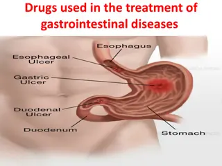
Chronic peptic ulcer Peptic Ulcer Disease Locations May occur in any portion of the GI tract exposed to acidic gastric juices 98% located in first portion of duodenum or stomach, ratio = 4:1 Esophagus . as a result of GERD or acid secretion by ectopic gastric mucosa. Gastric mucosa within a Meckel diverticulum can result in peptic ulceration of adjacent mucosa.
Peptic Ulcer Disease Gastric ulcers Pathophysiology The mucosal defences against acid attack consist of: 1. Mucus-bicarbonate barrier 2. The surface epithelium.
Peptic Ulcer Disease Gastric ulcers Pathophysiology The mucosal defences against acid attack consist of: 1. Mucus-bicarbonate barrier Duodeno-gastric reflux ( bile ) 2. The surface epithelium. 1. NSAIDs (blocking the synthesis of the prostaglandins) 2. H. pylori infection, ( cytotoxins and ammonia) Thus peptic ulcers in the stomach, breakdown of mucosal defence is much more important than excessive acid production.
Peptic Ulcer Disease Duodenal ulcers Pathophysiology Increased production of acid assumes more importance in the pathogenesis of duodenal ulceration H. pylori-infected individuals secrete 2-6 times as much acid as non-infected persons Helicobacter Pylori does not colonise normal duodenal epithelium. Helicobacter colonises areas of gastric metaplasia in duodenal ulceration because there is gastric metaplasia in response to excess acid. Gastric metaplasia paves the way for colonisation by Helicobacter + = Duodenal ulcers Increased production of acid Helicobacter P
Peptic Ulcer Disease Pathophysiology Duodeno-gastric reflux ( bile ) Hyperacidity Gastric ulcers Duodenal ulcers H. pylori NSAIDs H pylori infection of the pyloric antrum is present in nearly all patients with chronic duodenal ulcer and approximately 75% of patients with chronic gastric ulcer. Although more than 70% of individuals with PUD are infected by H. pylori, fewer than 20% of H. pylori infected individuals develop peptic ulcer.
H. pylori chronic active gastritis
Morphology Gross usually less than 20 mm in diameter but they may > 100 mm in diameter. The classic peptic ulcer is a round to oval, sharply punched-out defect In contrast, heaped-up margins are more characteristic of cancers Microscopy the base consists of necrotic tissue and polymorph exudate overlying inflamed granulation tissue which merges with mature fibrous (scar) tissue.
Clinical features Epigastric pain (the most common symptom) Gnawing or burning sensation Occurs 2-3 hours after meals Relieved by food or antacids Patient awakens with pain at night. Some present with complications such as iron deficiency anemia, frank hemorrhage, or perforation.
Complications Hemorrhage. Penetration. The ulcer penetrates the full thickness of the stomach or duodenal wall, progressing into adherent underlying tissue, particularly the pancreas or liver. Penetration of the pancreas often manifests clinically as severe back pain. Perforation. This leads to peritonitis. Fibrous stricture. This is seen in peptic ulcer of the esophagus and the stomach (pyloric stenosis) Malignant change. This is extremely uncommon.
Therapy Current therapies for PUD are aimed at 1. H. pylori eradication 2. Neutralization of gastric acid and acid suppression primarily with proton pump inhibitors and H2 blockers