Percutaneous SpyGlass Cholangioscopy for Intrahepatic Bile Duct Calculi Treatment
Bile duct injuries post laparoscopic cholecystectomy can lead to biliary strictures and recurrent cholangitis. Discover how Percutaneous SpyGlass Cholangioscopy is utilized for treating intrahepatic bile duct calculi, overcoming challenges in altered anatomy cases.

Download Presentation

Please find below an Image/Link to download the presentation.
The content on the website is provided AS IS for your information and personal use only. It may not be sold, licensed, or shared on other websites without obtaining consent from the author.If you encounter any issues during the download, it is possible that the publisher has removed the file from their server.
You are allowed to download the files provided on this website for personal or commercial use, subject to the condition that they are used lawfully. All files are the property of their respective owners.
The content on the website is provided AS IS for your information and personal use only. It may not be sold, licensed, or shared on other websites without obtaining consent from the author.
E N D
Presentation Transcript
Percutaneous SpyGlass Cholangioscopy For Treatment Of Intrahepatic Bile Duct Calculi Dr Melissa Wright1(BSc, MD); Dr Justin Chan1(MBBS, FRACS); Dr Ryan Rudolph2 (MBBCh, FRANZCR, EBIR); A/Prof Koroush S. Haghighi1(MBBS, FRACS, FACS). Affiliation: Prince of Wales Clinical School, University of New South Wales 1Department of General Surgery, Prince of Wales Hospital, Sydney, NSW 2Department of Medical Imaging, Prince of Wales Hospital, Sydney, NSW Published in ANZJS November 2021
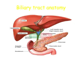
Background Bile duct injuries are a complication of laparoscopic cholecystectomy Long-term sequelae - biliary strictures, recurrent cholangitis. Retrieval of stones/dilatation of strictures in biliary systems that have had roux-en-y hepaticojejunostomy are challenging altered anatomy Generally percutaneous transhepatic cholangiography (PTC) required not amenable to standard endoscopic retrograde cholangiopancreatography (ERCP).
Novel technique allowing endoscopists to access and visualise small and otherwise difficult-to-reach bile ducts. Fibre-optic probe connected to a camera and inserted into a flexible catheter attached to the duodenoscope which is operated by a single endoscopist. Four-way tip deflection of the catheter allows for better visualisation of the biliary ducts. The system also allows treatment of calculi by electrohydraulic lithotripsy (EHL). Used in conjunction with ERCP for difficult cases of intraductal calculi and assessment of indeterminate biliary strictures requiring direct visualisation of the biliary tree. What is SpyGlass?
Case: 45F - laparoscopic cholecystectomy age 19 - complicated by a Bismuth Type E3 bile duct injury Roux-en-y reconstruction with a 60cm loop - complicated by a biliary leak/revision of the reconstruction. Developed extensive biliary stricture multiple dilatations over the next 5yrs Referred at 24yrs to our unit underwent further dilatations via PTC Well for the following 10yrs increasing frequency of cholangitis. Increasing number of intrahepatic calculi needed definitive intervention. Surgical exploration would have been challenging cholangioscopy via PTC
A) MRCP 2013 demonstrating dilated intra-hepatic ducts. B) MRCP 2021 prior to cholangioscopy intervention demonstrating numerous intra-hepatic calculi
Procedure: While patient was well PTC biliary drain inserted and balloon dilatation of the right hepatic anastomotic stricture performed. Cholangiogram demonstrated numerous filling defects throughout the right biliary tree consistent with intrahepatic calculi (Fig A) SpyGlass with EHL was performed calculi fragments were flushed through the anastomosis and a balloon sweep performed A) PTC Cholangiogram demonstrating multiple right biliary tree calculi and right hepatic duct anastomotic stricture B) Post-spyglass/lithotripsy cholangiogram demonstrating reduced calculi burden and patent ducts Cholangioscopy confirmed a significant reduction in calculi burden, however multiple residual fragments remained (Fig B)
Progress CT cholangiogram 2 weeks later demonstrated a filling defect in hepatic segment V. SpyGlass cholangioscopy/EHL procedure was repeated 4x total at two-weekly intervals Completion cholangiogram demonstrated a widely patent hepaticojejunostomy anastomosis and no sizeable intra-ductal calculi A digital subtraction angiography guided embolisation of the liver tract was performed and the PTC drain was removed.
Progress The patient was followed up two months post-procedure and had no evidence of residual calculi on MRCP and was asymptomatic
Discussion: Safe and effective option for treating biliary calculi - challenging anatomy post hepaticojejunal reconstruction. Not previously described in Australia Internationally, similar case reports demonstrate comparable results (1-5) - no large cohort studies have been published. A single-centre case series published in 2020 case series of four patients - concluding that it is a suitable technique for evaluating and treating biliary pathologies with challenging anatomy (6).
PTC-SpyGlass is a safe and effective treatment for intrahepatic calculi, in patients who have failed conventional approaches. Conclusion:
2 additional patients have undergone the same treatment with SpyGlass cholangioscopy and EHL awaiting 2 month follow up results to publish case series Case series in progress
References 1. Bhandari S, Bathini R, Sharma A, Maydeo A. Percutaneous endoscopic management of intrahepatic stones in patients with altered biliary anatomy: A case series. Indian Journal of Gastroenterology. 2016;35. 2. Du L, D'Souza P, Thiesen A, Girgis S, Owen R, McNally D, et al. Percutaneous transhepatic cholangioscopy for indeterminate biliary strictures using the SpyGlass system: a case series. Endoscopy. 2015;47(11):1054-6. 3. Raheel M, Dyer J, Curran F, Menon S. Cholangioscopy-guided electrohydraulic lithotripsy of a large bile duct stone through a percutaneous T-tube tract. VideoGIE. 2018;3. 4. Tellez-Avila F, Duarte-Medrano G, Andraca F, Gallardo-Cabrera V, Herrera Mora D. Percutaneous laser application using the SpyGlass system in a patient with intrahepatic lithiasis, liver cirrhosis, and surgically altered anatomy. Endoscopy. 2016;48:E49-E50. 5. Alabraba E, Travis S, Beckingham I. Percutaneous transhepatic cholangioscopy and lithotripsy in treating difficult biliary ductal stones: Two case reports. World J Gastrointest Endosc. 2019;11(4):298-307. 6. Tripathi N, Mardini H, Koirala N, Raissi D, Emhmed Ali SM, Frandah WM. Assessing the utility, findings, and outcomes of percutaneous transhepatic cholangioscopy with Spyglass(TM) Direct visualization system: a case series. Translational gastroenterology and hepatology. 2020;5:12.


 MRCP 2013 demonstrating dilated intra-hepatic ducts. B)")