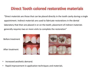
Discover the vital link between periodontal health and dental restoration for long-term success. Explore biologic considerations, esthetic factors, and occlusal considerations to ensure optimal outcomes. Learn about evaluating and correcting biologic width violations, as well as margin placement techniques to maintain periodontal health.
Please find below an Image/Link to download the presentation.
The content on the website is provided AS IS for your information and personal use only. It may not be sold, licensed, or shared on other websites without obtaining consent from the author. If you encounter any issues during the download, it is possible that the publisher has removed the file from their server.
You are allowed to download the files provided on this website for personal or commercial use, subject to the condition that they are used lawfully. All files are the property of their respective owners.
The content on the website is provided AS IS for your information and personal use only. It may not be sold, licensed, or shared on other websites without obtaining consent from the author.
BIOLOGIC CONSIDERATION ESTHETIC CONSIDERATIONS OCCLUSAL CONSIDERATIONS SPECIAL CONSIDERATIONS
BIOLOGIC CONSIDERATIONS Margin Placement & Biologic Width: Biologic Width: The C\T attachment occupies 1.07mm of space above the crest of alveolar bone &that the junctional epithelium below the base of the gingival sulcus ocupies another 0.97mm of space above the C\T attachment.
Correction of Biologic-Width Violation 1.SURGICALLY: By removing bone away from proximity to the restorative margins. Drawback: High risk of inter papillary recession. 2. ORTHODONTICALLY: By extruding the tooth out of the socket. Two Methods By applying low extrusive forces By rapid orthodontic extrusion
Margin Placement 1) Supragingival 2) Equigingival 3) Sub gingival Supra-Gingival Margin Placed in Non-esthetic area Least impact on periodontium Equi-gingival Margin At the crest of marginal gingiva
More impact on periodontium More plaque retentive-Gingival Inflammtion Subgingival margin Below the gingiva Greatest biologic risk May voilates the gingival attachment apparatus
restoration is extended sub-gingivally for For adequete resistance& retention To make significant contact & contour To mask the tooth-restoration interface gingivally. Too Sub-Gingivally located margins may results..... Unpredicted bone loss Gingival tissue recession
MARGIN PLACEMENT GUIDELINES i. If the sulcus probes 1.5mm or less,Place the margin 0.5mm below the gingival crest. ii. If sulcus probes more than 1.5mm- Place the margin 1\2 depth of the sulcus below the crest. iii. If sulcus probes more than 2.0mm, Esp. On facial aspect- Gingivectomy should be done & create a 1.5mm sulcus Apply Rule No. 1
CLINICAL PROCEDURE The Supra-gingival & Sub-gingival margins are very simple to place but the sub-gingival margin placement is rather difficult. So, Prior the placement of margin sub-gingivally the preparation should be extended to the free gingival margin facially & inter-proximally. The steps are as follows:
TISSUE RETRATION For protection from abrasion 2) For proper access BY GINGIVAL RETRACTION CORDS For Rule 1 Margin 2 Cords are used 1 cord is placed 0.5mm below the prepared margin. 2nd Cord is displaces the first cord apically & sits b\w the margin & tissue.
For Rule 2 Margin 1) 2 Large diameter Cords are used. Thick cord is placed for impression. 2) Electro-surgery is necessary to remove overhanging tissues.
This process: 1) Protects the tissues 2) Creates the correct axial reduction Establishes a desired sub-gingival level margin.
PROVISIONAL RESTORATION 3 Critical areas which must be appropriate to maintain the health & position of the Gingival tissues: 1. Marginal Fit 2. Contour 3.Surface finish
MARGINAL FIT Open margins can provide shelter for micro- organisms and may be responsible for inflammatory response.
CROWN CONTOUR Ideal contour provides access for hygiene & has fullness to cre create desired gingival form and good esthetics
. SUB-GINGIVAL DEBRIS Leaving debris below the tissue during the restorative procedure can cause adverse Pdl. Changes. Causes: Retraction cords Impression material Provisional materialCROWN CONTOUR Cements Temporary/permanent
ESTHETIC CONSIDERATIONS Inter-proximal Embrasure Form Any change in shape or form of embrassure Change in height & form of the papilla Food impaction, Accumulation of micro-organisms & Plaque accumulation
Too Wide Embrasure Flattened & Blunt Papilla Too Narrow Embrasure Inflammed Papilla Ideal Embrasure Healthy & pointed Papilla Restorative Correction of Open Gingival Embrasure 2 Causes: -In adequete papillary height due to bone loss -Highly located inter-proximal contact
This can be Corrected by Restorative Procedure 1)Direct Bonded Restoration Move the contact towards papilla 2) Indirect Restoration Contour & Embrassure form should be established in provisional restoration
Management of Gingival Embrassure form with Pdl. Recession Anterior Region: Carry the inter-proximal contact towards papilla In multiple units Porcelain Papillae on restoration using tissue coloured ceramic. Posterior Region: Contacts should be moved far enough apically.
Pontic Design 4 Types 1) Sanitary 2) Ridge-lap 3) Modified Ridge-lap 4) Ovate Ideal Form, Highly esthetic, Most Commonly used in Maxillary Anterior Region
OCCLUSAL CONSIDERATIONS Excessive occlusal load or trauma is a primary factor in Pocket formation, Gingival changes and Osseous & C\T destruction. Excessive occlusal force results in radiographic changes, Widening of Pdl. Spaces, Crestal funneling,Alteration in furcation bone quality & Variation in appearance of lamina dura.
SPECIAL CONSIDERATIONS Restoration of Root Resected Tooth One piece cast or core is indicated Development of appropriate contour for hygienic access Avoid any excess convexities
Splinting Apllied to: Bonded External Appliances Intra Coronal Appliances Indirect Cast Restorations Any inflammation of the Pdl,. Supporting tissues must be controlled before making a decision for splinting Bcoz Inflammation may cause mobility in presence of normal occlusal forces & Pdl. Support.
Anterior Aesthetic Surgery More Imp. In Anterior Region Gingivectomy, Apically displaced flaps with osseous recontouring & Use of Orthodontics in positioning the gingiva apically or coronally by Extruding or Intruding the teeth. Computer Imaging for Visual preview A Stone cast of patient s own teeth may be used Composite or Acrylic veneer is constructed on the cast extending gingivally in a correct position.
Pdl. Treatment before Restorative Management Sequence of Treatment: Extraction of Hopeless teeth Construction of Temp. Partial denture Periodontal Therapy is performed 2 month after Pdl. Therapy Relocate the margins Construct the Final Restoration
Phase 1 Therapy 1. Controlling active dental diseases 2.Pdl. Surgical Procedures: Crown lengthening surgery To prevent exposure of crown margin & Root surface after placement. Ridge augmentation: For correcting Ridge discrepancies 2 Techniques 1) The Roll Technique 2) The C\T Tissue Tunnel Technique