Phenotypic Classification of Leptospira Species
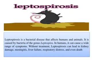
Leptospira species like L. interrogans and L. biflexa are classified based on their pathogenicity and serovars. They cause diseases like leptospirosis and are transmitted through zoonotic sources. Risk factors, epidemiology, and pathogenesis are important aspects to consider.

Download Presentation

Please find below an Image/Link to download the presentation.
The content on the website is provided AS IS for your information and personal use only. It may not be sold, licensed, or shared on other websites without obtaining consent from the author.If you encounter any issues during the download, it is possible that the publisher has removed the file from their server.
You are allowed to download the files provided on this website for personal or commercial use, subject to the condition that they are used lawfully. All files are the property of their respective owners.
The content on the website is provided AS IS for your information and personal use only. It may not be sold, licensed, or shared on other websites without obtaining consent from the author.
E N D
Presentation Transcript
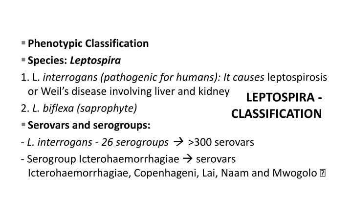
Phenotypic Classification Species: Leptospira 1. L. interrogans (pathogenic for humans): It causes leptospirosis or Weil s disease involving liver and kidney 2. L. biflexa (saprophyte) Serovars and serogroups: - L. interrogans - 26 serogroups >300 serovars - Serogroup Icterohaemorrhagiae serovars Icterohaemorrhagiae, Copenhageni, Lai, Naam and Mwogolo LEPTOSPIRA - CLASSIFICATION
Serogroups of Leptospira interrogans Australis Grippoty phosa Autumna lis adis Ballum Icteroha emorrha giae Bataviae Javanica Tarassovi Canicola Leptone ma Celledoni Lyme Cynopter Mini Sarmin Hebdom Sejroe LEPTOSPIRA - CLASSIFICATION Semaran ga Hurstbri dge Ranarum Turneria
Size: 612 m 0.1 m pass through filters used to sterilize culture medium Tightly & regularly coiled, with characteristic hooked ends - interrogans resembling interrogation mark Single endoflagellum attached at pole highly motile exhibiting spinning and translational movements Dark ground or phase contrast microscope Silver impregnation method and immunofluorescence LEPTOSPIRA INTERROGANS
Zoonotic Source: Rats, dogs, cattle and pigs Mode of transmission: Directhuman-to-human transmission does not occur - Indirect contact with water, moist soil and wet surfaces contaminated with animal urine - Direct contact with urine and products of parturition, placenta of infected animals Seasonality: Rainy and post monsoon period Epidemiology
Risk factors that promote transmission - Lower socioeconomic status, Urban and rural slum areas - Rainfall and floods - Occupational exposure: Agricultural workers, fishermen, sewer workers 3R s: Three important epidemiological determinants - rodents, rainfall and rice field Epidemiology
1. First phase (septicemic phase): Entry through mucosa/ abraded skin bloodstream disseminate brain, liver, lung, heart & kidney Vascular damage: capillaries, medium and large -sized vessels Penetration and invasion of tissues due to active motility and release of hyaluronidase Pathogenesis
2. Second phase (immune phase): - Antibodies develop, spirochetes disappear from the blood - Antigen antibody complexes are deposited in various organs Renal colonization - excreted in urine Clinical Manifestations - Incubation period 5 14 days 1. Mild anicteric febrile illness: (90%) - Biphasic; septicemic phase immune phase 2. Weil s disease (Hepato-renal- hemorrhagic syndrome): (10% ) - Severe form of icteric illness patients. Pathogenesis
Mild anicteric febrile illness First stage 3-10 days (septicemic) Fever, Myalgia, Headache, Second stage 10-30 days (immune) Meningitis, Uveitis, optic neuritis, chorioretinitis Rash, Fever Peripheral neuropathy Clinical Findings Leptospirosis Conjunctival, suffusion, Abdominal pain, Pharyngeal, erythema Without , Exudates, Vomiting From blood and CSF Absent Susceptible to antibiotics Isolation Serum IgM Antibiotics From Urine Present Refractory to treatment
Weils disease First stage 3-10 days (septicemic) Second stage 10-30 days (immune) Clinical Findings High grade fever, Liver- Jaundice and raised liver enzymes Leptospirosis Hemorrhages- Pulmonary hemorrhage, Petechiae & purpura, Conjunctival hemorrhage, Gastrointestinal hemorrhage Kidney- Raised serum urea and creatinine & Renal failure Blood and CSF Serum IgM Absent Antibiotics Susceptible to antibiotics Isolation Urine Present Refractory to treatment
Specimens: - First 10 days - CSF and blood urine - Between 10 and 30 days - Urine Microscopy: Wet films: Dark ground or phase contrast microscope Staining: sliver impregnation stains - Fontana stain & modified Steiner technique Laboratory Diagnosis
growing - Incubated at 30oC for 4 6 weeks at pH 7.2 7.5 - Culture fluid examined under dark ground microscope periodically EMJH medium (Ellinghausen, McCullough, Johnson, Harris) - albumin fatty acid supplement added to the basal media containing 0.1% agar. - Dinger s ring - Dense ring Isolation
- Korthof s medium with rabbit blood - Fletcher s semisolid medium Advantages - Isolation confirms the diagnosis - To maintain the stock culture Isolation
Disadvantages of Culture - Laborious, technically demanding and time-consuming - False-positive - contamination of culture media with other organisms or saprophytic leptospires False-negative - prior use of antibiotics, or incubating in improper temperature and pH Animal inoculation: Hamsters (4 6 weeks old) & young guinea pigs peritoneal fluid is examined for leptospires Laboratory Diagnosis
Genus-specific tests: uses antigen prepared from nonpathogenic L.biflexa Patoc 1 strain - Macroscopic slide agglutination test - Microcapsule agglutination test (MCAT) - Latex agglutination test - ELISA: IgM and IgG detected separately - Lepto dipstick assay: detects IgM Serology for antibody detection:
Serology for antibody detection: Immunochromatographic test (ICT): It detects IgM and IgG antibodies separately
Serovar-specific test: Microscopic agglutination test (MAT) - detects antibodies against specific serovars of L. Interrogans - Gold standard method and reference test Cross agglutination and absorption test (CAAT) detects relatedness between the strains Serology for antibody detection:
occurs - Target Genes -16S or 23S rRNA or IS1533 insertion sequence - PCR is not serovar-specific - PCR-RFLP or PFGE - to determine the genomo species Faine s criteria: WHO- approved guideline - based on clinical, epidemiological and laboratory findings Altered renal and liver Laboratory Diagnosis
Vaccine Whole cell vaccines (mono- or polyvalent) - Route Subcutaneous - Doses: 2 doses at 15-days interval third dose 4 6 month later biannual revaccination Efficacy rate - 60 100% Prevention
General Measures - Chemoprophylaxis - doxycycline - General sanitation approaches - proper waste disposal, Rodent control - Avoidance of swimming in contaminated places - Health education Prevention
REFRENCES Text Book Of Medical Microbiology by Ananth Narayan Paniker Text Book Of Medical Microbiology by D R Arora Text Book Of Medical Microbiology by A S Sastry Text Book Of Medical Microbiology by Baweja Text Book Of Medical Microbiology by Satish Gupte


:")