Physics Case Study: Bone Survey Image Quality Degradation Explained
In this physics case study, the possible cause of image quality degradation in a bone survey study is discussed. The comparison of lumbar spine images reveals the impact of factors such as source-to-image distance, field of view, and mAs settings on image quality. The diagnosis points to the use of a higher kV setting, affecting contrast in the images. Explore the implications of these technical aspects on radiographic images in this insightful analysis.
Download Presentation

Please find below an Image/Link to download the presentation.
The content on the website is provided AS IS for your information and personal use only. It may not be sold, licensed, or shared on other websites without obtaining consent from the author. If you encounter any issues during the download, it is possible that the publisher has removed the file from their server.
You are allowed to download the files provided on this website for personal or commercial use, subject to the condition that they are used lawfully. All files are the property of their respective owners.
The content on the website is provided AS IS for your information and personal use only. It may not be sold, licensed, or shared on other websites without obtaining consent from the author.
E N D
Presentation Transcript
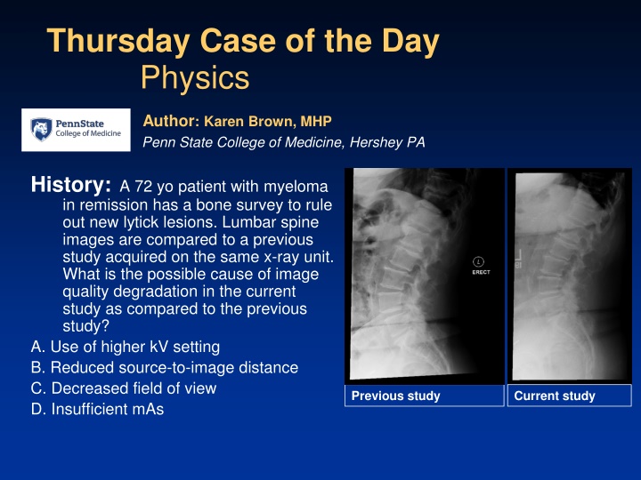
Thursday Case of the Day Physics Author: Karen Brown, MHP Penn State College of Medicine, Hershey PA History: A 72 yo patient with myeloma in remission has a bone survey to rule out new lytick lesions. Lumbar spine images are compared to a previous study acquired on the same x-ray unit. What is the possible cause of image quality degradation in the current study as compared to the previous study? A. Use of higher kV setting B. Reduced source-to-image distance C. Decreased field of view D. Insufficient mAs Previous study Current study
Findings: Reduced source-to-image distance would result in increased magnification in the current study as compared to the previous study. The current study does not appear to be magnified compared to the previous study. The field of view in the current study is slightly decreased compared to the previous study however use of tighter collimation (smaller field of view) would reduce the amount of scatter radiation produced resulting in improved image quality. Insufficient mAs would result in more quantum mottle (noise) which is not observed in the current study.
Diagnosis: A. Use of higher kV setting
Discussion: The previous study was acquired using 85 kV and 200 mAs. The current study was acquired using 95 kV and 310 mAs. The lower kV used in the previous study produced an image with greater contrast as compared to the current study. Previous study Current study
