Pituitary Hormones and Their Control by the Hypothalamus
The article delves into the intricate relationship between the pituitary hormones and their regulation by the hypothalamus. Dr. Noori M. Luaibi provides valuable insights into the mechanisms behind hormone control, shedding light on this vital aspect of endocrine physiology.

Download Presentation

Please find below an Image/Link to download the presentation.
The content on the website is provided AS IS for your information and personal use only. It may not be sold, licensed, or shared on other websites without obtaining consent from the author.If you encounter any issues during the download, it is possible that the publisher has removed the file from their server.
You are allowed to download the files provided on this website for personal or commercial use, subject to the condition that they are used lawfully. All files are the property of their respective owners.
The content on the website is provided AS IS for your information and personal use only. It may not be sold, licensed, or shared on other websites without obtaining consent from the author.
E N D
Presentation Transcript
Pituitary Pituitary Hormones and Their Hormones and Their Control by the Hypothalamus Control by the Hypothalamus DR. DR. NOORI NOORI M M LUAIBI LUAIBI
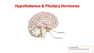
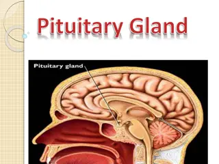
PITUITARY GLAND AND ITS RELATION TO THE HYPOTHALAMUS PITUITARY GLAND: TWO DISTINCT PARTS THE ANTERIOR AND POSTERIOR LOBES. The pituitary gland (Figure 75 1), also called the 1hypophysis, is a small gland about 1 centimeter in diameter and 0.5 to 1 gram in weight that lies in the sella turcica, a bony cavity at the base of the brain, and is connected to the hypothalamus by the pituitary (or hypophysial) stalk. Physiologically, the pituitary gland is divisible into two distinct portions: the anterior pituitary, also known as the adenohypophysis, and the posterior pituitary, also known as the neurohypophysis. Between these is a small, relatively avascular zone called the pars intermedia, which is almost absent in the human being but is much larger and much more functional in some lower animals. Embryologically, the two portions of the pituitary originate from different sources the anterior pituitary from Rathke s pouch, which is an embryonic invagination of the pharyngeal epithelium, and the posterior pituitary from a neural tissue outgrowth from the hypothalamus.Theorigin of the anterior pituitary from the pharyngeal epithelium explains the epithelioid nature of its cells, and the origin of the posterior pituitary from neural tissue explains the presence of large numbers of glial-type cells in this gland.
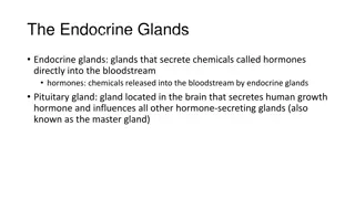
Six important peptide hormones plus several less important ones are secreted by the anterior pituitary, and two important peptide hormones are secreted by the posterior pituitary. THE HORMONES OF THE ANTERIOR PITUITARY PLAY MAJOR ROLES IN THE CONTROL OF METABOLIC FUNCTIONS THROUGHOUT THE BODY, (FIGURE 75 2), Growth hormone promotes growth of the entire body by affecting protein formation, cell multiplication, and cell differentiation. Adrenocorticotropin (corticotropin) controls the secretion of some of the adrenocortical hormones, which affect the metabolism of glucose, proteins, and fats. Thyroid-stimulating hormone (thyrotropin) controls the rate of secretion of thyroxine and triiodothyronine by the thyroid gland, and these hormones control the rates of most intracellular chemical reactions in the body. Prolactin promotes mammary gland development and milk production. Two separate gonadotropic hormones, follicle-stimulating hormone and luteinizing hormone, control growth of the ovaries and testes, as well as their hormonal and reproductive activities. THE TWO HORMONES SECRETED BY THE POSTERIOR PITUITARY PLAY OTHER ROLES. Antidiuretic hormone (also called vasopressin) controls the rate of water excretion into the urine, thus helping to control the concentration of water in the body fluids. Oxytocin helps express milk from the glands of the breast to the nipples during suckling and possibly helps in the delivery of the baby at the end of gestation.
ANTERIOR PITUITARY GLAND CONTAINS SEVERAL DIFFERENT CELL TYPES THAT SYNTHESIZE AND SECRETE HORMONES. Usually, there is one cell type for each major hormone formed in theanterior pituitary gland.With special stains attached to high-affinity antibodies that bind with the distinctive hormones, at least five cell types can be differentiated provides (Figure 75 3) a summary of these cell types, the hormones they produce, and their physiological actions. These five cell types are: 1. Somatotropes human growth hormone (hGH) 2. Corticotropes adrenocorticotropin (ACTH) 3. Thyrotropes thyroid-stimulating hormone (TSH) 4. Gonadotropes gonadotropic hormones, which include both luteinizing hormone (LH) and folliclestimulating hormone (FSH) 5. Lactotropes prolactin (PRL) About 30 to 40 per cent of the anterior pituitary cells are somatotropes that secrete growth hormone, and about 20 per cent are corticotropes that secrete ACTH. Each of the other cell types accounts for only 3 to 5 per cent of the total; nevertheless, they secrete powerful hormones for controlling thyroid function, sexual functions, and milk secretion by the breasts. Somatotropes stain strongly with acid dyes and are therefore called acidophils. Thus, pituitary tumors that secrete large quantities of human growth hormone are called acidophilic tumors.
POSTERIOR PITUITARY HORMONES ARE SYNTHESIZED BY CELL BODIES IN THE HYPOTHALAMUS. The bodies of the cells that secrete the posterior pituitary hormones are not located in the pituitary gland itself but are large neurons, called magnocellular neurons, located in the supraoptic and paraventricular nuclei of the hypothalamus. The hormones are then transported in the axoplasm of the neurons nerve fibers passing from the hypothalamus to the posterior pituitary gland.
HYPOTHALAMUS CONTROLS PITUITARY SECRETION Almost all secretion by the pituitary is controlled by either hormonal or nervous signals from the hypothalamus. Indeed, when the pituitary gland is removed from its normal position beneath the hypothalamus and transplanted to some other part of the body, its rates of secretion of the different hormones (except for prolactin) fall to very low levels. Secretion from the posterior pituitary is controlled by nerve signals that originate in the hypothalamus and terminate in the posterior pituitary. In contrast, secretion by the anterior pituitary is controlled by hormones called hypothalamic releasing and hypothalamic inhibitory hormones (or factors) secreted within the hypothalamus itself and then conducted, , (Figure 75 4), to the anterior pituitary through minute blood vessels called hypothalamic-hypophysial portal vessels. In the anterior pituitary, these releasing and inhibitory hormones act on the glandular cells to control their secretion.
The hypothalamus receives signals from many sources in the nervous system. Thus, when a person is exposed to pain, a portion of the pain signal is transmitted into the hypothalamus. Likewise, when a person experiences some powerful depressing or exciting thought, a portion of the signal is transmitted into the hypothalamus. Olfactory stimuli denoting pleasant or unpleasant smells transmit strong signal components directly and through the amygdaloid nuclei into the hypothalamus. Even the concentrations of nutrients, electrolytes, water, and various hormones in the blood excite or inhibit various portions of the hypothalamus. Thus, the hypothalamus is a collecting center for information concerning the internal well-being of the body, and much of this information is used to control secretions of the many globally important pituitary hormones.
HYPOTHALAMIC-HYPOPHYSIAL PORTAL BLOOD VESSELS OF THE ANTERIOR PITUITARY GLAND The anterior pituitary is a highly vascular gland with extensive capillary sinuses among the glandular cells. Almost all the blood that enters these sinuses passes first through another capillary bed in the lower hypothalamus. The blood then flows through small hypothalamic-hypophysial portal blood vessels into the anterior pituitary sinuses. (Figure 75 4) shows the lowermost portion of the hypothalamus, called the median eminence, which connects inferiorly with the pituitary stalk. Small arteries penetrate into the substance of the median eminence and then additional small vessels return to its surface, coalescing to form the hypothalamic-hypophysial portal blood vessels. These pass downward along the pituitary stalk to supply blood to the anterior pituitary sinuses.
HYPOTHALAMIC RELEASING AND INHIBITORY HORMONES ARE SECRETED INTO THE MEDIAN EMINENCE. Special neurons in the hypothalamus synthesize and secrete the hypothalamic releasing and inhibitory hormones that control secretion of the anterior pituitary hormones.These neurons originate in various parts of the hypothalamus and send their nerve fibers to the median eminence and tuber cinereum, an extension of hypothalamic tissue into the pituitary stalk. The endings of these fibers are different from most endings in the central nervous system, in that their function is not to transmit signals from one neuron to another but rather to secrete the hypothalamic releasing and inhibitory hormones into the tissue fluids. These hormones are immediately absorbed into the hypothalamic- hypophysial portal system and carried directly to the sinuses of the anterior pituitary gland.
HYPOTHALAMIC RELEASING AND INHIBITORY HORMONES CONTROL ANTERIOR PITUITARY SECRETION. The function of the releasing and inhibitory hormones is to control secretion of the anterior pituitary hormones. For most of the anterior pituitary hormones, it is the releasing hormones that are important, but for prolactin, a hypothalamic inhibitory hormone probably exerts more control. The major hypothalamic releasing and inhibitory hormones are the following: 1. Thyrotropin-releasing hormone (TRH), whichcauses release of thyroid- stimulating hormone 2. Corticotropin-releasing hormone (CRH), which causes release of adrenocorticotropin 3. Growth hormone releasing hormone (GHRH), which causes release of growth hormone, and growth hormone inhibitory hormone (GHIH), also called somatostatin, which inhibits release of growth hormone 4. Gonadotropin-releasing hormone (GnRH), which causes release of the two gonadotropic hormones, luteinizing hormone and follicle-stimulating hormone 5. Prolactin inhibitory hormone (PIH), which causes inhibition of prolactin secretion There are some additional hypothalamic hormones, including one that stimulates prolactin secretion and perhaps others that inhibit release of the anterior pituitary hormones
PHYSIOLOGICAL FUNCTIONS OF GROWTH HORMONE 1. Growth Hormone Promotes Growth of Many Body Tissues 2. Growth Hormone Has Several Metabolic Effects 3. Growth Hormone Promotes Protein Deposition in Tissues 4. Enhancement of Amino Acid Transport Through the Cell Membranes 5. Enhancement of RNA Translation to Cause Protein Synthesis by the Ribosomes. 6. Increased Nuclear Transcription of DNA to Form RNA. 7. Decreased Catabolism of Protein and Amino Acids. 8. Growth Hormone Enhances Fat Utilization for Energy 9. Ketogenic Effect of Growth Hormone. 10. Growth Hormone Decreases Carbohydrate Utilization 11. Necessity of Insulin and Carbohydrate for the Growth- Promoting Action of Growth Hormone. 12. Growth Hormone Stimulates Cartilage and Bone Growth 13. Growth Hormone Exerts Much of Its Effect Through Intermediate Substances Called Somatomedins (Also Called Insulin-Like Growth Factors )
REGULATION OF GROWTH HORMONE SECRETION For many years it was believed that growth hormone was secreted primarily during the period of growth but then disappeared from the blood at adolescence. This has proved to be untrue. After adolescence, secretion decreases slowly with aging, finally falling to about 25 per cent of the adolescent level in very old age. Growth hormone is secreted in a pulsatile pattern, increasing and decreasing. The precise mechanisms that control secretion of growth hormone are not fully understood, but several factors related to a person s state of nutrition or stress are known to stimulate secretion: (1) starvation, especially with severe protein deficiency; (2) hypoglycemia or low concentration of fatty acids in the blood; (3) exercise; (4) excitement; (5) trauma. (6) ghrelin, a hormone secreted by the stomach before meals ,
Table 753 FACTORS THAT STIMULATE OR INHIBIT SECRETION OF GROWTH HORMONE STIMULATE GROWTH HORMONE INHIBIT GROWTH HORMONE SECRETION 1. Decreased blood glucose 2. Decreased blood free fatty acid 3. Starvation or fasting, protein deficiency 4. Trauma, stress, excitement 5. Exercise 6. Testosterone, estrogen 7. Deep sleep ( stages II and IV) 8. Growth hormone releasing hormone Inhibit Growth Hormone Secretion Secretion 1. glucose Increased blood glucose 2. Increased blood free fatty acids 3. Aging 4. Obesity 5. Growth hormone inhibitory hormone (somatostatin) 6. Growth hormone (exogenous) 7. Somatomedins (insulin-like growth factors)
ABNORMALITIES OF GROWTH HORMONE SECRETION Panhypopituitarism. This term means decreased secretion of all the anterior pituitary hormones. The decrease in secretion may be congenital (present from birth), or it may occur suddenly or slowly at any time during life, most often resulting from a pituitary tumor that destroys the pituitary gland. Dwarfism. Most instances of dwarfism result from generalized deficiency of anterior pituitary secretion (panhypopituitarism) during childhood. In general, all the physical parts of the body develop in appropriate proportion to one another, but the rate of development is greatly decreased. A child who has reached the age of 10 years may have the bodily development of a child aged 4 to 5 years, and the same person at age 20 years may have the bodily development of a child aged 7 to 10 years. A person with panhypopituitarydwarfism does not pass through puberty and never secretes sufficient quantities of gonadotropic hormones to develop adult sexual functions. In one third of such dwarfs, however, only growth hormone is deficient; these persons do mature sexually and occasionally reproduce. In one type of dwarfism (the African pygmy and the L vi-Lorain dwarf), the rate of growth hormone secretion is normal or high, but there is a hereditary inability to form somatomedin C, which is a key step for the promotion of growth by growth hormone.
PANHYPOPITUITARISM IN THE ADULT. Panhypopituitarism first occurring in adulthood frequently results from one of three common bnormalities.Two tumorous conditions, craniopharyngiomas or chromophobe tumors, may compress the pituitary gland until the functioning anterior pituitary cells are totally or almost totally destroyed.Thethird cause is thrombosis of the pituitary blood vessels. This abnormality occasionally occurs when a new mother develops circulatory shock after the birth of her baby. The general effects of adult panhypopituitarism are (1) hypothyroidism, (2) depressed production of glucocorticoids by the adrenal glands, and (3) suppressed secretion of the gonadotropic hormones so that sexual functions are lost. Thus, the picture is that of a lethargic person (from lack of thyroid hormones) who is gaining weight (because of lack of fat mobilization by growth, adrenocorticotropic, adrenocortical, and thyroid hormones) and has lost all sexual functions. Except for the abnormal sexual functions, the patient can usually be treated satisfactorily by administering adrenocortical and thyroid hormones.
Gigantism. Occasionally, the acidophilic, growth hormone producing cells of the anterior pituitary gland become excessively active, and sometimes even acidophilic tumors occur in the gland. As a result, large quantities of growth hormone are produced. All body tissues grow rapidly, including the bones. If the condition occurs before adolescence, before the epiphyses of the long bones have become fused with the shafts, height increases so that the person becomes a giant up to 8 feet tall. The giant ordinarily has hyperglycemia, and the beta cells of the islets of Langerhans in the pancreas are prone to degenerate because they become overactive owing to the hyperglycemia. Consequently, in about 10per cent of giants, full-blown diabetes mellitus eventually develops. In most giants, panhypopituitarism eventually develops if they remain untreated, because the gigantism is usually caused by a tumor of the pituitary gland that grows until the gland itself is destroyed. This eventual general deficiency of pituitary hormones usually causes death in early adulthood. However, once gigantism is diagnosed, further effects can often be blocked by microsurgical removal of the tumor or by irradiation of the pituitary gland.
Acromegaly. If an acidophilic tumor occurs after adolescence that is, after the epiphyses of the long bones have fused with the shafts the person cannot grow taller, but the bones can become thicker and the soft tissues can continue to grow. Enlargement is especially marked in the bones of the hands and feet and in the membranous bones, including the cranium, nose, bosses on the forehead, supraorbital ridges, lower jawbone, and portions of the vertebrae, because their growth does not cease at adolescence. consequently, the lower jaw protrudes forward, sometimes as much as half an inch, the forehead slants forward because of excess development of the supraorbital ridges, the nose increases to as much as twice normal size, the feet require size 14 or larger shoes, and the fingers become extremely thickened so that the hands are almost twice normal size. In addition to these effects, changes in the vertebrae ordinarily cause a hunched back, which is known clinically as kyphosis. Finally, many soft tissue organs, such as the tongue, the liver, and especially the kidneys, become greatly enlarged.
POSTERIOR PITUITARY GLAND AND ITS RELATION TO THE HYPOTHALAMUS The posterior pituitary gland, also called the neurohypophysis, is composed mainly of glial-like cells called pituicytes.Thepituicytesdo not secrete hormones; they act simply as a supporting structure for large numbers of terminal nerve fibers and terminal nerve endings from nerve tracts that originate in the supraoptic and paraventricular nuclei of the hypothalamus,(Figure 75-9) These tracts pass to the neurohypophysis through the pituitary stalk (hypophysial stalk).The nerve endings are bulbous knobs that contain many secretory granules.Theseendings lie on the surfaces of capillaries, where they secrete two posterior pituitary hormones: 1. antidiuretic hormone (ADH), also called vasopressin 2. oxytocin If the pituitary stalk is cut above the pituitary gland but the entire hypothalamus is left intact, the posterior pituitary hormones continue to be secreted normally, after a transient decrease for a few days; they are then secreted by the cut ends of the fibers within the hypothalamus and not by the nerve endings in the posterior pituitary. The reason for this is that the hormones are initially synthesized in the cell bodies of the supraoptic and paraventricular nuclei and are then transported in combination with carrier proteins called neurophysins down to the nerve endings in the posterior pituitary gland, requiring several days to reach the gland.
ADH is formed primarily in the supraoptic nuclei, whereas oxytocin is formed primarily in the paraventricular nuclei. Each of these nuclei can synthesize about one sixth as much of the second hormone as of its primary hormone. When nerve impulses are transmitted downward along the fibers from the supraoptic or paraventricular nuclei, the hormone is immediately released from the secretory granules in the nerve endings by the usual secretory mechanism of exocytosis and is absorbed into adjacent capillaries. Both the neurophysin and the hormone are secreted together, but because they are only loosely bound to each other, the hormone separates almost immediately. The neurophysin has no known function after leaving the nerve terminals.
CHEMICAL STRUCTURES OF ADH AND OXYTOCIN Both oxytocin and ADH (vasopressin) are polypeptides, each containing nine amino acids. Their amino acid sequences are the following: Vasopressin: Cys-Tyr-Phe-Gln-Asn-Cys-Pro-Arg- GlyNH2 Oxytocin: Cys-Tyr-Ile-Gln-Asn-Cys-Pro-Leu-GlyNH2 Note that these two hormones are almost identical except that in vasopressin, phenylalanine and arginine replace isoleucine and leucineof the oxytocin molecule. The similarity of the molecules explains their partial functional similarities.
PHYSIOLOGICAL FUNCTIONS OF ADH The injection of extremely minute quantities of ADH as small as 2 nanograms can cause decreased excretion of water by the kidneys (antidiuresis). Briefly, in the absence of ADH, the collecting tubules and ducts become almost impermeable to water, which prevents significant reabsorption of water and therefore allows extreme loss of water into the urine, also causing extreme dilution of the urine. Conversely, in the presence of ADH, the permeability of the collecting ducts and tubules to water increases greatly and allows most of the water to be reabsorbed as the tubular fluid passes through these ducts, thereby conserving water in the body and producing very concentrated urine. The precise mechanism by which ADH acts on the collecting ducts to increase their permeability is only partially known. Without ADH, the luminal membranes of the tubular epithelial cells of the collecting ducts are almost impermeable to water. However, immediately inside the cell membrane are a large number of special vesicles that have highly waterpermeablepores called aquaporins.
When ADH acts on the cell, it first combines with membrane receptors that activate adenylyl cyclase and cause the formation of cAMP inside the tubular cell cytoplasm.This causes phosphorylation of elements in the special vesicles, which then causes the vesicles to insert into the apical cell membranes, thus providing many areas of high water permeability. All this occurs within 5 to 10 minutes. Then, in the absence of ADH, the entire process reverses in another 5 to 10 minutes. Thus, this process temporarily provides many new pores that allow free diffusion of water from the tubular fluid through the tubular epithelial cells and into the renal interstitial fluid.Water is then absorbed from the collecting tubules and ducts by osmosis.
OXYTOCIC HORMONE Oxytocin Causes Contraction of the Pregnant Uterus. The hormone oxytocin, in accordance with its name, powerfully stimulates contraction of the pregnant uterus, specially toward the end of gestation. Therefore, many obstetricians believe that this hormone is at least partially responsible for causing birth of the baby. This is supported by the following facts: 1. In a hypophysectomized animal, the duration of labor is prolonged indicating a possible effect of oxytocin during delivery. 2. The amount of oxytocin in the plasma increases during labor, especially during the last stage. 3. Stimulation of the cervix in a pregnant animal elicits nervous signals that pass to the hypothalamus and cause increased secretion of oxytocin.
OXYTOCIN AIDS IN MILK EJECTION BY THE BREASTS. Oxytocin also plays an especially important role in lactation a role that is far better understood than its role in delivery. In lactation, oxytocin causes milk to be expressed from the alveoli into the ducts of the breast so that the baby can obtain it by suckling. This mechanism works as follows: The suckling stimulus on the nipple of the breast causes signals to be transmitted through sensory nerves to the oxytocin neurons in the paraventricularand supraoptic nuclei in the hypothalamus, which causes release of oxytocin by the posterior pituitary gland. The oxytocin is then carried by the blood to the breasts, where it causes contraction of myoepithelial cells that lie outside of and form a latticework surrounding the alveoli of the mammary glands. In less than a minute after the beginning of suckling, milk begins to flow. This mechanism is called milk letdown or milk ejection.