Plant Tissue Systems and Functions in Botany Study
Explore the fundamentals of plant tissue systems including simple and complex tissues derived from meristematic tissues. Learn about the three major plant organs - stem, root, and leaves, and their tissue compositions, as well as the classification and characteristics of plant tissues. Discover the roles of epidermal, ground, and vascular tissue systems in plant physiology.

Download Presentation

Please find below an Image/Link to download the presentation.
The content on the website is provided AS IS for your information and personal use only. It may not be sold, licensed, or shared on other websites without obtaining consent from the author. If you encounter any issues during the download, it is possible that the publisher has removed the file from their server.
You are allowed to download the files provided on this website for personal or commercial use, subject to the condition that they are used lawfully. All files are the property of their respective owners.
The content on the website is provided AS IS for your information and personal use only. It may not be sold, licensed, or shared on other websites without obtaining consent from the author.
E N D
Presentation Transcript
Presented By Garima Yadav Research Scholar Mohanlal Sukhadia University, Udaipur Department Of Botany Mohanlal Sukhadia University, Udaipur Department Of Botany
Forceps, Dissecting needles, Glass droppers, Good and sharp razor, Safety blade, A fine hair brush Dissecting microscope, Compound microscope Slides Cover glasses Watch glasses, Petri dishes Fast green( soft tissues) Safranin( hard tissues)
Transverse Longitudinal (I) Radial Longitudinal section (R.L.s) (ii) Tangential Longitudinal section (T.L.s.)
Mount with glycerin drop on slide and cover with cover slip and observe under microscope Transfer the section into watch glass containing safranin Transfer the section into watch glass containing water Wash with water and stain with fast green Section cutting
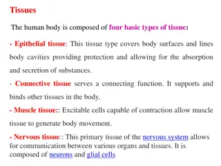
Three fundamental organs of plant are - stem, root and leaves. All these organs are made of tissue systems. The tissues constituting the tissue system are derived from meristematic tissues. These are situated only at a few places called meristems. The tissues are classified into two major groups simple tissues and the complex tissues. The simple tissues are made of only one type cells. These include parenchyma, sclerenchyma, collenchyma, etc. The complex tissues are made of more than one type of cells. Complex tissues include xylem and phloem. The xylem consists of cell types such as tracheids, vessels, xylem fibers and xylem parenchyma. The phloem is composed of sieve cells , companion cells, phloem_ fibers and phloem parenchyma.
Groups of some of these different types of tissues together perform a common function and are called tissue system. There are three tissue systems epidermal tissue system, ground or fundamental tissue system and vascular tissue system. Tissue is a group of cells which may be similar or (simple tissue) or dissimilar in character (complex tissue) and is specialized for a particular function. Characteristics functions and distribution of some of these plant tissues are given in the following table.
Tissue Living or dead WaIl material Cellulose, pectins and hemicellulos es Cell Shape Usually isodiamet -ric, sometime -s elongated Main functions Distribution Parenchyma Living Packing tissue. Supporting herbaceous plants ,Metabolically active. Intercellular air spaces allow gaseous exchange. Food storage. Transport of materials through cells or cell walls. Cortex, pith, medullary rays and packing tissue in xylem and phloem.
2. Modified parenchyma Living Cellulose, pectins and hemicelluloses , and covering of cutin Elongated and flattened Protection from desiccation and infection. Hairs and glands may have additional functions. Single layer of cells covering entire primary plant body. (a) Epidermis (b) Mesophyll Living Cellulose, pectins and hemicelluloses Isodiametric chlorenchyma, irregular or column shaped depending on location Photosynthesis (contains chloroplasts). Storage of starch. Between upper and lower epidermis of leaves.
Living Cellulose, pectins and hemicellulose s, and deposits of suberin (Casparian strips) As epidermis Selective barrier to movement of water and mineral salts (between cortex and xylem) in roots. Starch sheath with possible role in geotropic response in stems. Around vascular tissue. (innermost layer of cortex) (e) Endodermis (d) Pericycle Living Cellulose, pectins and hemicellulose -s As parenchyma In roots, it retains meristematic activity producing lateral roots and contributing to secondary growth if this occurs. In roots between central vascular tissue and endodermis.
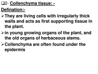
3.Collenchyma Living Cellulose, pectins and hemicellulose s Elongated and polygonal with tapering ends Support (a mechanical function). Outer regions of cortex, e.g. angles of stems, midrib of leaves. 4. Sclerenchyma Mainly lignin. Cellulose pectins and hemi- celluloses also present. Elongated and polygonal with tapering interlocking ends. Support (purely mechanical). Outer regions of cortex, pericyc1e of stems, xylem and phloem a) Fibers Dead b) Selereids As fibers Roughly isodiametric, though variations occur Support or mechanical protection Cortex, pith, phloem, shells and stones of fruits, seed coats. Dead
5. Xylem. Mixture of living and dead cells. Xylem also contains fibers and parenchyma (a) Tracheids Dead Mainly lignin. Cellulose, pectins and hemicellul -oses also present. Mainly lignin. Cellulose, pectins and hemicellul oses also present. Elongated and tubular Translocat -ion of water and mineral salts. Support. Vascular system (b) Vessels. Dead Elongated but broad and cylindrical Translocat ion of water and mineral salts. Support. Vascular system
5. Phloem. Mixture of living and dead cells. (a) Sieve tubes. Living Cellulose , pectins and hemicell- uloses. Cellulose , pectins and hemicell- uloses. Elongate- d and tubular Transloc ation of organic solutes (food) Work in associati on with sieve tubes Vascular system (b) Compani on cells Living Elongate- d and narrow Vascular system
Characters 1. Endodermis Stems Mayor may not be present. Conjoint, collateral and endarch. Roots Always very clearly marked Radial and exarch. 2. Vascular bundles
Characters Dicotyledons Monocotyledons 1. Hypodermis May or may not be present, if present mostly collenchymatous Generally present, sclerenchymatous. 2. Cortex A few layers of parenchyma extend up to the endodermis 3. Endodermis Generally absent; mostly represented by endodermoid cells; If present, in the form of a ring The cells following hypodermis are not differentiated. They are generally parenchymatous and extend from hypodermis up to the centre of the axis. It is known as ground tissue.
4. Pericycle Present between the vascular tissue and cortex; either parenchymatous or sclerenchymatous, one to few layered. s. Medullary ray A strip of parenchyma between the vascular bundles. 6. Pith A central and well marked out cylinder present; parenchymatous or sclerenchymatous Well marked pith can not be distinguished 7. Vascular bundles (a) Conjoint, collaterall bicollateral endarch, open. (b) Arranged in a ring. (c) Almost all of them are uniform In size. (d) Phloem parenchyma present. (e) Bundle sheath absent. (a) Conjoint, collateral, endarch and closed. (b) Scattered throughout ground tissue. (c) Larger toward the centre and Smaller outside. (d) Phloem parenchyma absent. (e) Well developed bundle sheath present.
Characters Dicotyledons Monocotyledons 1. Vascular bundles There are about 2-6 protoxylem groups (i.e. condition is diarch to hexarch); rarely more groups. The number of protoxylem groups generally exceeds 2-6, therefore, the condition is polyarch; rarely only a few. 2. Pericycle It generally gives rise to lateral roots, vascular cambium and cork cambium. Only lateral roots are produced. 3. Cambium It appears later to form a complete ring between the xylem and phloem groups. It is altogether absent. 4. Pith Small or absent. Large and well developed.
Primary growth is growth that occurs as a result of cell division at shoot apical meristem and root apical meristem, causing them to elongate, and gives rise to primary tissue.
In dicotyledons, the stem and the root both grow once again after the plant acquires primary structure. This growth is known as secondary growth. ,It occurs due to the activity of a secondary meristem or two lateral meristems- The vascular cambium and the phellogen or cork cambium. Vascular cambium forms secondary xylem and secondary phloem. Phellogen originates in the extrastelar region and forms periderm. The plants become woody and the girth increases. The tissues added by the vascular cambium are secondary xylem (wood) and secondary phloem Many dicotyledons and monocotyledons, however show the different type of growth from the normal type described above, termed as anomalous or abnormal growth.
Scattered vascular bundles in dicotyledons Vascular bundles arranged in rings in monocotyledons. Cortical bundles. Medullary bundles Intraxylary or internal phloem. Separate xylem and phloem bundle. Absence of vessels in xylem. Ploystelic condition.
Abnormal behavior of normal cambium. Abnormal behavior of abnormal cambium. Formation of accessory cambial rings. Formation of interxylary phloem.
Outline appears almost circular in transverse section. Epidermis. 1. It is an outermost single layer of cells. 2. The outer face has thick cuticle. Cortex. 1. It is many layers deep. The region is differentiated into outer collenchymas and inner parenchyma. 2. Collenchymas follows epidermis. It is three to five cells deep. The walls the neighboring cells are thickened. 3. Parenchyma follows the zone of collenchymas. It forms rest of the cortex. It contains numerous chloroplasts. Intercellular spaces are present. Endodermis and pericycle. These layers are indistinct. Vascular tissue system. 1. There' are many vascular bundles which are arranged in rings. A zone of secondary tissues is also very distinct.
2. The outermost ring has many bundles. Due to secondary growth, phloem occurs in the form of crushed and obliterated patches. Abundant prosenchyma (conjunctive tissue) is present. Secondary phloem forms a complete ring. Cambium that follows separates phloem and xylem. The primary xylem groups are situated close to the pith. Protoxylem is endarch and the vascular bundles are conjoint, collateral, endarch and open. 3. The innermost ring consists of two vascular bundles. Each bundle is conjoint, collateral, endarch and open. The bundles lie close to the pith, and are, therefore, known as medullary bundles. These bundles produce a small amount of secondary phloem and secondary xylem in radial rows. 4. The middle ring consists of six or seven (upto fourteen) bundles. These bundles are smaller than those of the inner ring. Each bundle is conjoint, collateral, endarch and open. Pith. In the centre a small parenchymatous pith is present.
Identification 1. Stem. Vascular bundles conjoint, collateral and open. 2. Dicotyledonous stem- 1. Cortex well differentiated. 2. Presence of secondary growth. 3. Vascular bundles in a ring.
1. Medullary bundles. The vascular bundles are arranged in three rings. Out of these, two inner rings occur in the pith and are, therefore, known as medullary bundles. The medullary (intrafascicular) cambium and produce a little amount of secondary tissues. 2. Abnormal secondary growth. In Boerhaavia, vascular bundles of the outermost ring have fascicular cambium. Later, interfascicular cambium also develops, thus forming a complete ring. The cambial ring produces secondary xylem and secondary phloem. However, this cambial ring soon stops functioning. A new ring (accessory cambium) appears later in the region of the pericycle. This ring of cambium, also, as in earlier cases functions only for some time. Such many (upto twenty two) accessory cambia are produced successively, much farther away into the cortex every time. This results in the formation of successive alternate zones of secondary xylem and secondary phloem. These rings or zones (of secondary tissues) are sometimes eccentrically developed. Cambia produce a very large amount of prosenchyma into which xylem remains scattered and at times becomes indistinguishable from it. bundles possess fascicular