Positron Emission Tomography (P.E.T) - A Comprehensive Guide

Positron Emission Tomography (P.E.T) is a noninvasive imaging technique that measures metabolic activity in cells. Learn about the history of positrons, how P.E.T works, its uses in detecting cancer, assessing heart conditions, and more. Discover the role of positrons, production process, benefits, and limitations of P.E.T in this detailed seminar submission

Download Presentation

Please find below an Image/Link to download the presentation.
The content on the website is provided AS IS for your information and personal use only. It may not be sold, licensed, or shared on other websites without obtaining consent from the author.If you encounter any issues during the download, it is possible that the publisher has removed the file from their server.
You are allowed to download the files provided on this website for personal or commercial use, subject to the condition that they are used lawfully. All files are the property of their respective owners.
The content on the website is provided AS IS for your information and personal use only. It may not be sold, licensed, or shared on other websites without obtaining consent from the author.
E N D
Presentation Transcript
www.studymafia.org Seminar On Positron Emission Tomography (P.E.T) Submitted To: Submitted By: www.studymafia.org www.studymafia.org
Content What is PET A little history about the positron What is a Positron? What happens after the positron is obtained? Uses How does it work? How is it performed? What are the benefits vs. risks? Things to consider Limitations
What is PET PET is a noninvasive, diagnostic imaging technique for measuring the metabolic activity of cells in the human body. It was developed in the mid 1970s and it was the first scanning method to give functional information about the brain.
A little history about the positron Existence first postulated in 1928 by Paul Dirac First observed in 1932 by Carl D. Anderson, who gave the positron its name. He also suggested to rename the electron to negatron but he was unsuccessful.
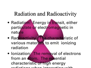
What is a Positron? A Positron is an anti-matter electron, it is identical in mass but has an apposite charge of +1. Positron can come from different number of sources, but for PET they are produced by nuclear decay. Nuclear decay is basically when unstable nuclei are produced in a cyclotron by bombarding the target material with protons, and as a result a neutron is released. 18-O + proton => 18-F + neutron In PET the target material is chosen so that the product of the bombardment decays to a more stable state isotope by emitting a positron, for instance 18-F has too many protons, so one of these protons decays into a neutron emitting in the process a positron an a neutrino. proton (+1 charge) => neutron (0 charge) + positron (+1 charge) + neutrino (0 charge) After decay, we re left with 18-O
What happens after the positron is obtained? Left over energy from the nuclear decay process is shared between the positron and the departing neutrino. Kinetic energy. Because of conservation of energy and momentum the positron is forced to stay and thus become useful. Positron begins its activity in colliding with other particles and gradually losing its kinetic energy and thus slowing down.
Uses Detect cancer. Determine whether a cancer has spread in the body. Assess the effectiveness of a treatment plan, such as cancer therapy. Determine if a cancer has returned after treatment. Determine blood flow to the heart muscle. Determine the effects of a heart attack, or myocardial infarction, on areas of the heart. Identify areas of the heart muscle that would benefit from a procedure such as angioplasty or coronary artery bypass surgery (in combination with a myocardial perfusion scan). Evaluate brain abnormalities, such as tumors, memory disorders and seizures and other central nervous system disorders. To map normal human brain and heart function.
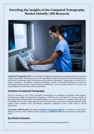
How does it work? Before the examination begins, a radioactive substance is produced in a machine called a cyclotron and attached, or tagged, to a natural body compound, most commonly glucose, but sometimes water or ammonia. Once this substance is administered to the patient, the radioactivity localizes in the appropriate areas of the body and is detected by the PET scanner. Different colors or degrees of brightness on a PET image represent different levels of tissue or organ function. For example, because healthy tissue uses glucose for energy, it accumulates some of the tagged glucose, which will show up on the PET images. However, cancerous tissue, which uses more glucose than normal tissue, will accumulate more of the substance and appear brighter than normal tissue on the PET images.
How is it performed? A nurse or technologist will take you into a special injection room, where the radioactive substance is administered as an intravenous injection (although in some cases, it will be given through an existing intravenous line or inhaled as a gas). It will then take approximately 30 to 90 minutes for the substance to travel through your body and accumulate in the tissue under study. During this time, you will be asked to rest quietly and avoid significant movement or talking, which may alter the localization of the administered substance. After that time, scanning begins. This may take 30 to 45 minutes. Some patients, specifically those with heart disease, may undergo a stress test in which PET scans are obtained while they are at rest and again after undergoing the administration of a pharmaceutical to alter the blood flow to the heart. Usually, there are no restrictions on daily routine after the test, although you should drink plenty of fluids to flush the radioactive substance from your body.
What are the benefits vs. risks? Because PET allows study of body function, it can help physicians detect alterations in biochemical processes that suggest disease before changes in anatomy are apparent with other imaging tests, such as CT or MRI. Because the radioactivity is very short-lived, your radiation exposure is low. The substance amount is so small that it does not affect the normal processes of the body. PET imaging has been shown to improve detection of a variety of cancers, and earlier tests have suggested this technique may be useful in identifying small tumors in patients with paraneoplastic neurological disorders. The radioactive substance may expose radiation to the fetus in patients who are pregnant or the infants of women who are breast- feeding. The risk to the fetus or infant should be considered in relation to the potential information gain from the result of the PET examination. If you are pregnant, you should inform the PET imaging staff before the examination is performed.
Things to consider You will remain still for a long time. Claustrophobic persons may feel some anxiety. Even though you may feel the desire to feel something due to the radioactivity, you will be disappointed, unless they mistakenly inject you plutonium gas.
Limitations PET can give false results if a patient's chemical balances are not normal. Specifically, test results of diabetic patients or patients who have eaten within a few hours prior to the examination can be adversely affected because of blood sugar or blood insulin levels. Also, because the radioactive substance decays quickly and is effective for a short period of time, it must be produced in a laboratory near the PET scanner. It is important to be on time for the appointment and to receive the radioactive substance at the scheduled time. PET must be done by a radiologist who has specialized in nuclear medicine and has substantial experience with PET. Most large medical centers now have PET services available to their patients. Medicare and insurance companies cover many of the applications of PET, and coverage continues to increase. Finally, the value of a PET scan is enhanced when it is part of a larger diagnostic work-up. This often entails comparison of the PET scan with other imaging studies, such as CT or MRI.
Reference www.google.com www.wikipedia.com www.studymafia.org