Postoperative Management Following Pituitary Surgery Overview

Pituitary lesions are common, with various presentations including mass effects and hormonal imbalances. Surgery is often the preferred treatment for different adenomas and tumors. Decision-making for resection should involve a multidisciplinary approach. Transsphenoidal surgery is the primary method, increasingly performed endoscopically. The efficacy of endoscopic versus microscopic views in tumor removal and remission rates for functional adenomas remains under investigation.

Uploaded on Apr 16, 2025 | 0 Views
Download Presentation

Please find below an Image/Link to download the presentation.
The content on the website is provided AS IS for your information and personal use only. It may not be sold, licensed, or shared on other websites without obtaining consent from the author.If you encounter any issues during the download, it is possible that the publisher has removed the file from their server.
You are allowed to download the files provided on this website for personal or commercial use, subject to the condition that they are used lawfully. All files are the property of their respective owners.
The content on the website is provided AS IS for your information and personal use only. It may not be sold, licensed, or shared on other websites without obtaining consent from the author.
E N D
Presentation Transcript
POSTOPERATIVE MANAGEMENT FOLLOWING PITUITARY SURGERY ACE July 2015 Dr M.Heidarpour
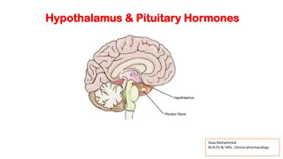
INTRODUCTION Pituitary lesions are common in the general population with approximately 15 to 20% of individuals having evidence of an identifiable lesion on brain imaging . As recently documented in the Central Brain Tumor Registry of the United States, pituitary tumors are the second most common brain pathology observed.
Pituitary lesions can present with mass effects (headaches, visual loss, cranial nerve dysfunction), pituitary hormonal hypersecretion or hormonal deficiency.
Although medical therapy is typically considered first line for prolactinomas, surgery is generally considered the treatment of choice for other endocrine-active pituitary adenomas (acromegaly, Cushing disease, and thyrotrope adenomas), as well as clinically nonfunctioning adenomas, pituitary apoplexy, Rathke cleft cysts, craniopharyngiomas, and other parasellar tumors such as meningiomas and clival chordomas.
The decision to proceed with neurosurgical tumor resection can be quite complex and is best considered in a multidisciplinary clinical setting. Once the decision has been made to consider postoperative management of the patient. Although a minority of patients with giant adenomas and/or invasive suprasellar adenomas may require a craniotomy, the vast majority (over 95%) of pituitary adenomas are best treated by transsphenoidal surgery (TSS).
Over the last 15 years, TSS is increasingly being performed using an endonasal endoscopic technique, often in a collaborative effort including a neurosurgeon and otolaryngologist .
While there is some evidence that the more panoramic endoscopic view leads to higher rates of complete tumor removal than the traditional microscopic view . It remains to be proven whether the endoscopic approach results in higher remission rates for functional adenomas such as those causing acromegaly or Cushing disease.
Postoperative management following tumor removal utilizing any approach or technique includes: 1. immediate inpatient assessment 2. short-term assessment for pituitary hormone recovery or deficits. 3. development of a long-term surveillance plan for tumor/lesion recurrence and endocrine status.
PREOPERATIVE EVALUATION All patients with sellar/suprasellar lesions Require: 1. medical history 2. physical exam including formal neuro- ophthalmologic assessment with formal visual field testing if the MRI shows evidence that the tumor is abutting or compressing the optic chiasm. 3. evaluation for potential medical comorbidities.
4. functioning tumor (e.g., prolactinoma, acromegaly, Cushing disease, or the rarely encountered thyrotropin-secreting tumor) 5.Hypopituitarism prior to development of the treatment plan.
Typical preoperative baseline endocrine testing includes: measurement of serum prolactin assessment of hypercortisolism if there is clinical suspicion of Cushingnsyndrome measurement of insulin-like growth factor-1 (IGF-I) with further assessment of growth hormone (GH) hypersecretion for acromegaly as indicated.
Preoperative prolactin measurements are essential, as medical therapy with dopamine agonists is the preferred initial treatment modality for prolactinoma patients . Sample dilution may be required in patients with large tumors and minimally elevated prolactin levels to rule out the hook effect that can be seen in some laboratory assays, leading to artificially low prolactin levels.
Assessment of pituitary hypofunction includes 1. hypothyroidism (thyroid-stimulating hormone and free thyroxine) hypogonadism (serum testosterone in males; follicle-stimulating hormone/luteinizing hormone, estradiol in a premenopausal woman with history of oligomenorrhea/ amenorrhea or postmenopausal females) 2. adrenal insufficiency (there are a number of protocols used including measurement of morning serum cortisol and cosyntropin stimulation testing). 3.
It is imperative that adrenocortical and thyroid status be evaluated prior to surgery to initiate appropriate hormonal replacement pre- and perioperatively if a deficiency is identified. It is important to recognize adrenal insufficiency preoperatively to ensure glucocorticoid coverage during surgery. It is likely important to identify hypothyroidism, as replacement may prevent potential surgical complications (i.e., cardiac dysfunction, hyponatremia, postoperative ileus)
Although found far less common in pituitary adenoma patients at initial presentation, diabetes insipidus (DI) can occur, particularly in concert with suprasellar lesions such as craniopharyngiomas, and needs to be identified and managed preoperatively. A careful history, along with measurement of serum electrolytes and urine specific gravity, will often alert the clinician to this condition.
Ideally, all patients will receive replacement hormonal therapy for adrenal insufficiency, hypothyroidism or DI if indicated prior to surgery. however, it may not be prudent to delay surgery to achieve normal replacement levels (i.e., hypothyroidism) in all cases. Initiation of sex steroid and GH replacement therapy is typically deferred until a later point in the postoperative management.
patients are generally evaluated for other potential comorbidities including diabetes mellitus and cardiopulmonary disease that should be addressed prior to surgery. Patients with acromegaly and Cushing disease are particularly prone to have associated cardiovascular and metabolic comorbidities such as: diabetes mellitus, hypertension, and cardiac dysfunction and may benefit from preoperative medical management.
There is debate regarding preoperative treatment of acromegaly or Cushing disease with hormone- lowering medications prior to surgical intervention. Currently, there is no consensus on the preoperative treatment of these diseases to improve surgical cure rate, but there may be a role in improving comorbidities and reducing complications.
PERIOPERATIVE MANAGEMENT .All patients with adrenal insufficiency identified preoperatively should be treated with stress-dose glucocorticoid treatment both peri- and postoperatively, with postoperative testing for endogenous production only if there is likelihood for a return to normal hypothalamicpituitary-adrenal axis function.
For patients with normal preoperative adrenal function: 1. glucocorticoids may be administered perioperatively to cover for potential iatrogenic adrenal insufficiency. 2. However, protocols in many centers involve steroid sparing management both peri- and postoperatively to avoid unnecessary exposure to glucocorticoids if possible.
Preoperative glucocorticoids in cushing disease Preoperative glucocorticoid replacement is not necessary unless cortisol production has been blocked by adrenal enzyme inhibitors. In this instance, the patient should be treated like a patient with adrenal insufficiency. .uptodate
Practice patterns are variable: Some surgeons do not administer glucocorticoids. 1. others give higher than normal glucocorticoid replacement intraoperatively and for one to three days postoperatively to avoid symptoms and signs of acute steroid withdrawal . There have been no comparisons of the benefits of one or the other approach. One typical regimen is dexamethasone 0.5 mg every six hours for four doses only, eg, only 24 hours of glucocorticoid therapy. .uptodate 2.
Perioperative glucocorticoid therapy entails virtually no risk for the patient, except that it must be stopped for 24 hours before serum cortisol can be measured to assess cure. Glucocorticoid replacement can be held for a few days, with careful observation for the development of adrenal insufficiency. .uptodate
Other complications, apart from surgically- related morbidity, include venous thrombosis and infection, which occurred in four and one patients, respectively. Since the risk of thromboembolic complications is increased in Cushing's syndrome, perioperative prophylaxis seems warranted in patients who are not ambulatory within a few days of surgery . .uptodate
POSTOPERATIVE MANAGEMENT The general framework for postoperative management of patients undergoing pituitary tumor surgery can be considered to occur in 2 main phases: early postoperative period longer term outpatient follow-up period, * initial outpatient management * long-term observation
Early Postoperative Management The postoperative inpatient stay typically lasts from 1 to 3 nights . Serious complications following pituitary adenoma surgery are relatively uncommon, particularly with an experienced surgical team.
TSS patients warrant careful monitoring for at least the first postoperative night and then typically in a monitored bed. At many pituitary centers, Foley catheters are removed after surgery, and arterial lines are not routinely used. More invasive monitoring may be used selectively for patients with significant medical comorbidities or those with large or complex parasellar lesions (e.g., giant or highly invasive pituitary adenomas, craniopharyngiomas, and meningiomas).
Major complications in the immediate postoperative period after TSS can be categorized into surgical and endocrine. The most serious early surgical complications(typically seen within 24-48 hours of surgery) include sellar hematoma, often with associated visual loss, diplopia, and/or headache and cerebrospinal fluid (CSF) leak.
Hydrocephalus, meningitis, sinusitis, and epistaxis may develop within the first 1 to 3 weeks after surgery. Patients should be monitored closely for potential neurologic or ophthalmologic deterioration using serial visual field assessments and neurologic exam, particularly in the first 24 hours following surgery.
An early postoperative CT or sellar MRI should be performed in any patient with a new or worsened neurological deficit 1. visual deterioration or diplopia, 2. significant rhinorrhea 3. suspected CSF leak.
Although not performed routinely at all centers, sellar imaging within 24 hours of surgery can also be useful to assess: skull base reconstruction integrity degree of pneumocephalus and for the presence of a sellar or suprasellar hematoma particularly after removal of larg macroadenoma, craniopharyngioma, or suprasellar meningioma.
The most important potential endocrine complications in the immediate postoperative period include: 1. fluid and electrolyte abnormalities 2. acute adrenal insufficiency Alterations in sodium and fluid balance are relatively common in the early postoperative phase. These include alterations in arginine vasopressin/antidiuretic hormone (ADH), either insufficiency causing central DI or excess leading to the syndrome of inappropriate ADH release (SIADH).
DI,which may occur in up to 25% of patients after pituitary adenoma surgery, is most frequently observed in within the first 48 hours of surgery . It is thought to occur from manipulation, traction, or disruption of the pituitary stalk during tumor removal leading to interruption of ADH release. Patients typically complain of excessive thirst and demonstrate polydipsia with polyuria and dilute urine(urine specific gravity typically 1.005), indicating an inability to concentrate the urine due to ADH deficiency.
Although usually transient, DI may be permanent if there is stalk transection . Fluid intake, urinary output, volume status, as well as serial serum sodium and urine specific gravity or urine osmolality should be assessed to allow prompt recognition of the condition.
One study showed a lower incidence of postoperative DI in patients who were not routinely treated with glucocorticoids . DI can be managed with desmopressin (DDAVP), which is available as a subcutaneous or intravenous (0.5-2 mcg every 24 hours as needed), intranasal (10 mcg metered dose), or oral formulation (often starting with 0.1-0.2 mg oral as a single dose with doses up to 0.3 mg oral 3 times daily sometimes necessary). Intranasal DDAVP is not generally used until after the nose has healed and nasal congestion has improved.
Many patients do not require any therapy as long as they are able to drink to thirst and their serum sodium remains within the normal range. For others, only 1 or 2 DDAVP doses may be needed as an inpatient before the condition resolves.
DI can be transient, permanent, or remit and then recur later postoperatively in a classic triphasic response . In the latter scenario, patients can initially develop DI in the first 24 to 48 hours followed by transient SIADH developing 4 to 10 days postoperatively, followed by the return of DI in a matter of weeks. When DI returns as the third phase, this disturbance can be permanent.
Management depends on the phase, and it is important not to overtreat the first phase of DI, which can result in severe hyponatremia during the potential subsequent SIADH phase. Fortunately, permanent DI occurs in only approximately 2% of patients following TSS.
Between 5 and 9% of patients may also develop isolated SIADH leading to symptomatic and delayed hyponatremia. Therefore, a sodium level is often checked 5 to 8 days postoperatively, typically after the patient has been discharged from the hospital.
Mild hyponatremia (i.e., 130-135 mmol/L) may be managed by fluid restriction as an outpatient. severe or symptomatic hyponatremia (typically <125 mmol/L) requires hospitalization for more aggressive management including use of fluid restriction, hypertonic saline or administration of vaptans, competitive antagonists of vasopressin receptors.
In a recent study by Jahangiri et al , postoperative hyponatremia was noted in 19% of subjects. In this study, use of vaptans resulted in more rapid plasma sodium correction versus hypertonic saline. but did lead to overcorrection in 1 subject. Therefore, selective use of vaptans in the setting of severe hyponatremia may be useful and potentially lead to shorter hospital stays.
Journal of Neuroscience Nursing
Finally, the clinician should be aware of the critical need to monitor adrenal function and replace glucocorticoids as needed following pituitary surgery.
There are several approaches toward glucocorticoid management in the peri- and postoperative timeframes. ranging from glucocorticoid treatment at surgery with gradual tapering steroid sparing protocols with careful observation
1.Some clinicians choose to empirically treat all patients perioperatively with glucocorticoids at stress doses (for example, 50-100 mg intravenous hydrocortisone) immediately prior to or during the operation followed by a gradual tapering schedule with reassessment of adrenal function during the early postoperative follow-up as necessary once on lower replacement doses.
2.Another approach is to administer pre- or intraoperative stress dose glucocorticoids only if the preoperative assessment suggests or confirms adrenal insufficiency. In a steroid-sparing protocol, perioperative steroids would be withheld entirely in a patient with normal preoperative adrenal function. In the days following surgery, early morning cortisol levels are measured and glucocorticoids initiated if the value is consistent with insufficiency.
measurement of a basal morning cortisol level is only suggestive of and not diagnostic of adrenal insufficiency. various cut points have been proposed, below which glucocorticoid replacement should be considered. Proposed cut points for guidance in management are based on studies that suggest a high likelihood of adrenal insufficiency with a morning cortisol less than 4 to 5 mcg/ dL and low likelihood if the level is above 10 to 15 mcg/dL.
There is no one best approach, and many factors need to be considered for an individual patient, but minimizing unnecessary glucocorticoid exposure may be desired. If glucocorticoids are initiated in the perioperative period based on morning cortisol levels dropping below a specified cut point, then physiologic replacement therapy should be maintained until further provocative testing (approximately 6 weeks postoperatively) can be performed to assess long-term replacement needs.
patients should be treated with 100 mg of hydrocortisone beginning at the induction of anesthesia. The dose should be gradually decreased during the next few days. 1. We recommend a replacement dose (eg, 15 to 25mg/day) following discharge until the initial postoperative evaluation four to six weeks after discharge. 2. Others recommend measuring serum cortisol on the third postoperative day, 24 hours after the previous dose of hydrocortisone, and if the value is low (less than 4mcg/dL [110 nmol/L]) or borderline (5 to 17 mcg/dL [138 to 469 nmol/L]), prescribing replacement hydrocortisone on discharge. uptodate


")