Process of Fertilization: Phases and Mechanisms Explained
Explore the intricate details of the fertilization process, from sperm movement to oocyte penetration. Learn about capacitation, acrosome reaction, and the fusion of gametes during the first week of development. Understand the phases of fertilization, including corona radiata and zona pellucida penetration, leading to the fusion of oocyte and sperm cell membranes. Dive into the fascinating journey of male and female gametes coming together for the formation of new life.

Download Presentation

Please find below an Image/Link to download the presentation.
The content on the website is provided AS IS for your information and personal use only. It may not be sold, licensed, or shared on other websites without obtaining consent from the author. If you encounter any issues during the download, it is possible that the publisher has removed the file from their server.
You are allowed to download the files provided on this website for personal or commercial use, subject to the condition that they are used lawfully. All files are the property of their respective owners.
The content on the website is provided AS IS for your information and personal use only. It may not be sold, licensed, or shared on other websites without obtaining consent from the author.
E N D
Presentation Transcript
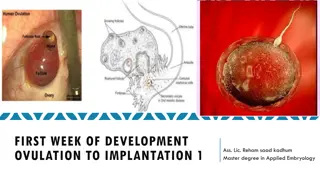
FIRST WEEK OF DEVELOPMENT BY: MBBSPPT.COM
FERTILIZATION Fertilization, the process by which male and female gametes fuse, occurs in the ampullary region of the uterine tube. This is the widest part of the tube and is close to the ovary. Spermatozoa may remain viable in the female reproductive tract for several days. Only 1% of sperm deposited in the vagina enter the cervix, where they may survive for many hours.
FERTILIZATION Movement of sperm from the cervix to the uterine tube occurs by muscular contractions of the uterus and uterine tube and very little by their own propulsion. The trip from cervix to oviduct can occur as rapidly as 30 minutes or as slow as 6 days. After reaching the isthmus, sperm become less motile and cease their migration. Spermatozoa are not able to fertilize the oocyte immediately upon arrival in the female genital tract but must undergo (1) capacitation and (2) the acrosome reaction to acquire this capability.
1. Capacitation is a period of conditioning in the female reproductive tract that in the human lasts approximately 7 hours. Thus, speeding to the ampulla is not an advantage, since capacitation has not yet occurred and such sperm are not capable of fertilizing the egg. 2. The acrosome reaction, which occurs after binding to the zona pellucida, is induced by zona proteins. This reaction release of enzymes needed to penetrate the zona pellucida, including acrosin- and trypsin-like substances.
THE PHASES OF FERTILIZATION INCLUDE: Phase 1: Penetration of the Corona Radiata Of the 200 to 300 million spermatozoa normally deposited in the female genital tract, only 300 to 500 reach the site of fertilization. Capacitated sperm pass freely through corona cells.
Phase 2: Penetration of the Zona Pellucida The head of the sperm is capped by an organelle c/a Acrosome (containing a trypsin-like protein digesting enzyme & hyaluronidase). When the head of sperm comes in contact with zona pellucida, an acrosome reaction is induced by the zona protein----The acrosome release digestive enzyme (acrosin & pepsin-like substance)---cause lysis of the zona pellucida and plasma membrane around the head of the sperm.---allows sperm to penetrate through the zona pellucida ---once the sperm penetrate the properties of zona pellucida change (zona reaction)---occurs that makes it impermeable to other sperms.
Phase 3: Fusion of the Oocyte and Sperm Cell Membranes The plasma membranes of sperm and oocyte come in contact and breakdown at the site of fusion. The head and tail of sperm enter the cytoplasm of the oocyte but its plasma membrane and mitochondrial sheath are left behind on the oocyte surface.---as soon as the sperm enters the oocyte a calcium wave appears in the cytoplasm of oocyte---that makes oocyte membrane impermeable to other sperms.
Figure-A Oocyte immediately after ovulation, showing the spindle of the second meiotic division. B. A spermatozoon has penetrated the oocyte, which has finished its second meiotic division. Chromosomes of the oocyte are arranged in a vesicular nucleus, the female pronucleus. Heads of several sperm are stuck in the zona pellucida. C. Male & female pronuclei. D,E. Chromosomes become arranged on the spindle, split longitudinally, and move to opposite poles. F. Two-cell stage.
CLEAVAGE Once the zygote has reached the two-cell stage, it undergoes a series of mitotic divisions, increasing the numbers of cells. These cells, which become smaller with each cleavage division, are known as blastomeres. Until the eight-cell stage, they form a loosely arranged clump. After the third cleavage, blastomeres maximize their contact with each other, forming a compact ball of cells held together by tight junctions. Approximately 3 days after fertilization, cells of the compacted embryo divide again to form a 16-cell morula(mulberry).
BLASTOCYST FORMATION About the time the morula enters the uterine cavity, fluid begins to penetrate through the zona pellucida into the intercellular spaces of the inner cell mass. Gradually, the intercellular spaces become confluent, and finally, a single cavity, the blastocele, forms.
At this time, the embryo is a blastocyst. Cells of the inner cell mass, now called the embryoblast. The outer cell mass are called trophoblast. Trophoblast: Flatten and form the epithelial wall of the blastocyst. The zona pellucida has disappeared, allowing implantation to begin.