Explore the world of protein homology modelling, ab initio structure prediction, and structural genomics to understand the intricacies of protein structure determination and its significance in biology. Learn about the steps involved, pitfalls to avoid, and the latest advancements in the field.
Please find below an Image/Link to download the presentation.
The content on the website is provided AS IS for your information and personal use only. It may not be sold, licensed, or shared on other websites without obtaining consent from the author. If you encounter any issues during the download, it is possible that the publisher has removed the file from their server.
You are allowed to download the files provided on this website for personal or commercial use, subject to the condition that they are used lawfully. All files are the property of their respective owners.
The content on the website is provided AS IS for your information and personal use only. It may not be sold, licensed, or shared on other websites without obtaining consent from the author.
After this lesson you should be able to: Explain the individual steps involved in calculating a protein homology model. Identify suitable templates for modelling. Outline the principles behind ab initio protein structure prediction. Describe the differences between homology modelling and ab initio structure prediction. Describe the major pitfalls in protein modelling. LEARNING OBJECTIVES
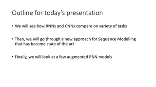
Protein homology modelling Individual steps Caveats Pitfalls Ab initio protein structure prediction Threading True ab initio methods OUTLINE
Why Do We Need Homology Modelling? Ab Initioprotein folding ( random sampling): 100 aa, 3 conf./residue gives approximately 1048 different overall conformations! Random sampling is NOT feasible, even if conformations can be sampled at picosecond (10-12 sec) rates. Levinthal s paradox Do homology modelling instead.
The structure of a protein is uniquely determined by its amino acid sequence (but sequence is sometimes not enough): prions pH, ions, cofactors, chaperones Structure is conserved much longer than sequence in evolution. Structure > Function >> Sequence HOW IS IT POSSIBLE?
There are currently ~47000 structures in the PDB (but only ~4000 if you include only ones that are not more than 30% identical and have a resolution better than 3.0 ). An estimated 25% of all sequences can be modeled and structural information can be obtained for ~50%. HOW OFTEN CAN WE DO IT?
Complete genomes Signaling proteins Disease-causing organisms Model organisms Membrane proteins Protein-ligand interactions WORLDWIDE STRUCTURAL GENOMICS Fold space coverage
10 year $600 million project initiated in 2000, funded largely by NIH. AIM: structural information on 10000 unique proteins (now 4-6000), so far 1000 have been determined. Improve current techniques to reduce time (from months to days) and cost (from $100.000 to $20.000/structure). 9 research centers currently funded (2005), targets are from model and disease-causing organisms (a separate project on TB proteins). STRUCTURAL GENOMICS IN NORTH AMERICA
Search with sequence Blast Psi-Blast Fold recognition methods Use biological information Functional annotation in databases Active site/motifs TEMPLATE IDENTIFICATION
1 2 3 4 5 6 7 8 9 10 11 12 13 14 PHE ASP ILE CYS ARG LEU PRO GLY SER ALA GLU ALA VAL CYS PHE ASN VAL CYS ARG THR PRO --- --- --- GLU ALA ILE CYS PHE ASN VAL CYS ARG --- --- --- THR PRO GLU ALA ILE CYS F 6 -3 2 0 D -2 0 C -3 -2 2 -2 -2 0 -2 2 -2 -1 2 I R P -2 -3 -1 -2 -3 -2 0 -2 -2 0 2 -1 -1 -1 0 L G S A E A V C -3 F N V C R T P E A I C 0 1 -1 0 0 -2 -2 5 -2 -2 -2 -2 -2 5 -1 0 0 1 -1 0 -1 -1 -2 -3 2 -2 0 0 -3 -2 -2 8 -2 -3 0 -1 -2 -1 -1 1 -3 5 -2 -2 2 -2 1 0 0 1 1 1 5 5 1 1 5 -1 -3 0 -2 -2 -2 -2 -1 -1 -2 -1 2 -2 -3 -3 -2 -1 -2 -3 -2 -2 8
1 2 3 4 5 6 7 8 9 10 11 12 13 14 PHE ASP ILE CYS ARG LEU PRO GLY SER ALA GLU ALA VAL CYS PHE ASN VAL CYS ARG THR PRO --- --- --- GLU ALA ILE CYS PHE ASN VAL CYS ARG --- --- --- THR PRO GLU ALA ILE CYS F 6 -3 2 0 -3 -2 -2 8 -2 -2 -2 -2 5 -2 0 0 -2 0 -2 -3 0 -3 2 -2 -3 0 -2 0 -1 -2 -1 -1 1 0 -3 5 -2 -2 2 -3 -2 -2 8 D -2 0 C -3 -2 2 -2 -2 0 -2 2 -2 -1 2 I P -2 -3 -1 -2 -3 -2 0 -2 -2 0 2 -1 -1 -1 0 -2 -3 -3 -2 -1 -2 -3 -2 -2 8 -1 0 0 1 -1 0 0 0 -1 2 -2 8 0 0 -2 1 0 1 0 1 -2 -2 -1 -1 -2 -1 2 -2 -3 -3 -2 -1 -2 -3 -2 -2 8 R L G S A E A V C -3 F N V C R T P E A I C 0 1 -1 0 0 -2 -2 5 -2 -1 0 0 1 1 5 -1 -1 -2 0 0 1 -1 -3 1 -1 -3 5 0 1 1 5 1 -1 -2 -2
1 2 3 4 5 6 7 8 9 10 11 12 13 14 PHE ASP ILE CYS ARG LEU PRO GLY SER ALA GLU ALA VAL CYS PHE ASN VAL CYS ARG THR PRO --- --- --- GLU ALA ILE CYS PHE ASN VAL CYS ARG --- --- --- THR PRO GLU ALA ILE CYS IMPROVING THE ALIGNMENT From Professional Gambling by Gert Vriend http://www.cmbi.kun.nl/gv/articles/text/gambling.html
Selecting the best template is crucial! The best template may not be the one with the highest % id (best p-value ) Template 1: 93% id, 3.5 resolution Template 2: 90% id, 1.5 resolution TEMPLATE QUALITY
Knowledge based: Searches PDB for fragments that match the sequence to be modelled (Levitt, Holm, Baker etc.). Energy based: Uses an energy function to evaluate the quality of the loop and minimizes this function by Monte Carlo (sampling) or molecular dynamics (MD) techniques. Combination LOOP MODELLING
Find fragments (10 per amino acid) with the same sequence and secondary structure profile as the query sequence. Combine them using a Monte Carlo scheme to build the loop. David Baker et al. LOOPS THE ROSETTA METHOD
Side chain rotamers are dependent on backbone conformation. Most successful method in CASP6 was SCWRL by Dunbrack et al.: Graph-theory knowledge based method to solve the combinatorial problem of side chain modelling. http://dunbrack.fccc.edu/SCWRL3.php PREDICTING SIDE CHAIN CONFORMATIONS
Prediction accuracy is high for buried residues, but much lower for surface residues Experimental reasons: side chains at the surface are more flexible. Theoretical reasons: much easier to handle hydrophobic packing in the core than the electrostatic interactions, including H-bonds to waters. SIDE CHAINS - ACCURACY
Energy minimization Molecular dynamics Big errors like atom clashes can be removed, but force fields are not perfect and small errors will also be introduced keep minimization to a minimum or matters will only get worse. REFINEMENT
If errors are introduced in the model, they normally can NOT be recovered at a later step The alignment can not make up for a bad choice of template. Loop modeling can not make up for a poor alignment. If errors are discovered, the step where they were introduced should be redone. ERROR RECOVERY
Most programs will get the bond lengths and angles right. The Ramachandran plot of the model usually looks pretty much like the Ramachandran plot of the template (so select a high quality template). Inside/outside distributions of polar and apolar residues can be useful. Biological/biochemical data Active site residues Modification sites Interaction sites VALIDATION
ProQ is a neural network based predictor that based on a number of structural features predicts the quality of a protein model. ProQ is optimized to find correct models in contrast to other methods which are optimized to find native structures. VALIDATION PROQ SERVER Arne Elofssons group: http://www.sbc.su.se/~bjorn/ProQ/
ProCheck http://www.biochem.ucl.ac.uk/~roman/procheck/procheck.html WhatIf server http://swift.cmbi.kun.nl/WIWWWI/ STRUCTURE VALIDATION
Eva-CM performs continuous and automated analysis of comparative protein structure modeling servers A current list of the best performing servers can be found at: http://cubic.bioc.columbia.edu/eva/doc/intro_cm.html HOMOLOGY MODELLING SERVERS
Threading and pair potentials Ab initio methods Human intervention (what kind of knowledge can be used for alignment and selection of templates?) Meta-servers (the principle, 3d jury) Summary of take-home messages OUTLINE
Example: Pair potentials How normal is it to observe a pair of an alanine and a valine separated by 20 residues in the sequence and 3 in space? (X) Compares a given sequence against known structures (folds). By using potentials that describe tendencies observed in known protein structures. How normal is it to observe any pair of residues separated by 20 residues and 3 in space? (Y) THREADING AND PAIR POTENTIALS Potential: log (X/Y)
Alignment score from structural fitness (pair potential) Deletions 7 4 6 2 5 8 10 9 3 1 How well does K fit environment at P6? If P8 is acidic then fine, if P8 is basic then poor .. A T N L Y K E T L .. POTENTIALS OF MEAN FORCE
Problem: No protein is average Interactions in proteins cannot only be described by pairs of amino acids The information in the potentials is partly captured with sequence profiles Today mostly used in HYBRID approaches in combination with profile-profile based methods Potentials can be used to score models based on different templates or alignments THREADING METHODS TODAY
Aim is to find the fold of native protein by simulating the biological process of protein folding. A VERY DIFFICULT task because a protein chain can fold into millions of different conformations. Use it only when no detectable homologues are available. Methods can also be useful for fold recognition in cases of extremely low homology (e.g. convergent evolution). AB INITIO METHODS
Rosetta method of the Baker group: Submit sequence to a number of secondary structure predictors. Compare fragments of 3 and 9 residues to library from know structures. Link fragments together. FRAGMENT-BASED AB INITIO MODELLING Use energy minimization techniques (Monte Carlo optimization) to calculate tertiary structure.
Use of energy potentials for scoring and computing models. Potentials should make models more native-like . These can be based on contact potentials, solvation potentials, Van der Waals repulsion and attractive forces, hydrogen bond potentials. Globularity/radius of gyration (ab initio). POTENTIALS FOR FINDING GOOD MODELS
Fragments with correct local structure PROBLEMS WITH EMPIRICAL POTENTIALS
Knowledge of function The best methods use maximum knowledge of query proteins. Cysteines forming disulfide bridges or binding e.g. zinc molecules Proteolytic cleavage sites Other metal binding residues Antibody epitopes or escape mutants Ligand binding Specialists can help to find a correct Results from CD or fluorescence experiments HUMAN INTERVENTION template and correct alignments. Knowledge of secondary structure
Democratic modeling The highest score hit is often wrong. Many prediction methods have the correct fold among the top 10- 20 hits. If many different prediction methods all have some fold among the top hits, this fold is probably correct. META-SERVERS
3DJury http://bioinfo.pl/meta/ Inspired by Ab initio modeling methods Average of frequently obtained low energy structures is often closer to the native structure than the lowest energy structure Find most abundant high scoring model in a list of prediction from several predictors Use output from a set of servers 1. Superimpose all pairs of structures 2. Similarity score based on # of C pairs within 3.5 3. Similar methods developed by A. Elofsson (Pcons) and D. Fischer (3D shotgun). EXAMPLE OF A META-SERVER
Because it is a meta-server it can be slow. If queue is too long some servers are skipped. Output is only C coordinates. What to do with the rest of the structure? Use e.g. maxsprout server to build sidechains and backbone atoms. http://www.ebi.ac.uk/maxsprout/ 3DJURY
Hybrid methods using both threading methods and profile-profile alignments are the best. Use only Ab initio methods if necessary and know that the quality is really low! Try to use as much knowledge as possible for alignment and template selections in difficult cases. Use meta-servers when you can. SUMMARY AB INITIO METHODS


 – initial alignment")


 with the same")