In this report, the structures of human tetrameric hemoglobin (PDB code: 3onz) and alcohol dehydrogenase (PDB code: 3tn7) are analyzed. Details include the resolution, chain information, and sequence comparisons. The report provides insights into the structural characteristics of these proteins.
Please find below an Image/Link to download the presentation.
The content on the website is provided AS IS for your information and personal use only. It may not be sold, licensed, or shared on other websites without obtaining consent from the author. If you encounter any issues during the download, it is possible that the publisher has removed the file from their server.
You are allowed to download the files provided on this website for personal or commercial use, subject to the condition that they are used lawfully. All files are the property of their respective owners.
The content on the website is provided AS IS for your information and personal use only. It may not be sold, licensed, or shared on other websites without obtaining consent from the author.
E N D
Presentation Transcript
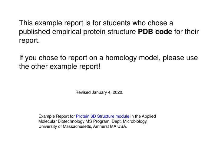
This example report is for students who chose a published empirical protein structure PDB code for their report. If you chose to report on a homology model, please use the other example report! Revised January 4, 2020. Example Report for Protein 3D Structure module in the Applied Molecular Biotechnology MS Program, Dept. Microbiology, University of Massachusetts, Amherst MA USA.
You may prepare your report slides in any of the following: Microsoft Powerpoint Google Slides Libre Office (please save as .pptx file) Open Office (please save as .pptx file) Powerpoint and Microsoft Office are free for UMass students. The other three are free for everyone. In my opinion, Google Slides is a little easier to use than Powerpoint, yet very similar. To access it, go to slides.google.com, login (or create a free account), and click the + box to start a new presentation. When your presentation report is finished and ready to submit for grading, email your .pptx file as an attachment (or a link to it in dropbox), or a link to your Google Slides report, to martzeric@yahoo.com.
Section 1: Name, PDB codes Your NAME here please Your EMAIL address here please Faculty name for your research lab here please Subject of your project here please I chose*: 3onz HUMAN TETRAMERIC HEMOGLOBIN: PROXIMAL NITRITE LIGAND AT BETA Function: Oxygen transport Resolution: 2.09 R = 0.218 Rfree = 0.280 WORSE THAN AVERAGE at this resolution I was assigned: 3tn7 Alcohol dehydrogenase. Enzyme that oxidizes alcohols to aldehydes or ketones. Resolution: 1.68 R = 0.146 Rfree = 0.181 Reliability: Better than average at this resolution * If you chose a homology model, use the other demo report!
Section 2A, Number of chains for 3onz Protein: 2 chains (2 distinct) DNA: 0 chains RNA: 0 chains Chain A: HEMOGLOBIN SUBUNIT ALPHA 141 residues (1 missing) of Protein. Source: Homo sapiens (Human). Other_details: Blood. Chain B: HEMOGLOBIN SUBUNIT BETA 146 residues (12 missing) of Protein. Source: Homo sapiens (Human). Other_details: Blood. Each chain is given a distinct color ( Cartoon view).
Section 2B, Number of chains for assigned model 3tn7 Protein: 2 chains (sequence identical) DNA: 0 chains RNA: 0 chains 2 Sequence-Identical Chains A, B: SHORT-CHAIN ALCOHOL DEHYDROGENASE 257 residues (including 23 missing) of Protein. Source: Thermococcus sibiricus. Expression system: Escherichia coli. Each chain is given a distinct color ( Cartoon view).
Section 3A: Full length vs. crystallized sequence for 3onz Chain A: Conclusions for chain B: Crystal: 141 amino acids Full length: 142 amino acids Chain B: Crystal: 146 amino acids Full-length: 147 amino acids Only the N-terminal Met was removed before crystallization. From 2-147 the sequences are identical. Nothing was added before crystallization. Below, full length vs. crystallized sequence alignment for chain B:
Section 3B: Full length vs. crystallized sequence for 3tn7 Chains A=B: Crystal: 257 amino acids Full length: 234 amino acids Conclusions: None of the full-length sequence was removed before crystallization. A 23-residue poly-histidine tag was added to the N-terminus to facilitate purification. Googling ssglvprgshmle reveals that LVGRPS is a thrombin cleavage site. From 24-257 the sequences are identical.
Section 4: Non-covalent interactions for 3onz Top: hydrophobic van der Waals interaction between two carbon atoms. Bottom: histidine nitrogen interacting with negatively charged oxygen (carboxyl). Hydrogen bond since His N epsilon has a hydrogen to donate. The Heme oxygen lacks a hydrogen, so must be the acceptor. Chain A Phe 46 Heme His 45 His 45 Note: If you use the built-in save static image in FirstGlance, the dashed distance lines will be almost invisible. Instead, capture your own snapshot from FirstGlance following these instructions but DO NOT turn on high quality .
Section 5A - Biological unit Asymmetric unit: 2 chains Biological unit: 4 chains Note: when saving these images, it is important to CANCEL hiding the text in JSmol. This preserves the identities of the molecules shown (upper left), which confirms which is the asymmetric unit, and which the biological unit ( MM1 ).
Section 5B - Biological unit for 3tn7. Asymmetric unit: 2 chains Biological unit: 4 chains Note: when saving these images, it is important to CANCEL hiding the text in JSmol. This preserves the identities of the molecules in the upper left, which confirms which is the asymmetric unit, and which the biological unit ( MM1 ).
Section 6, 3onz - Evolutionary Conservation http://consurf.tau.ac.il/results/1368001022/output.php Top: Lys 61, high conservation is unexpected. Bottom: Lys 66, high conservation is expected because it forms salt bridges with the carboxyls on the heme ligand.