Protein Structure
Proteins, essential in biological processes, display diverse functions shaped by their intricate 3D structures. Understanding amino acids as protein building blocks sheds light on their polymers and distinct chemical properties. Discover the crucial link between protein structure and function illustrated through enzymes catalyzing reactions, transport proteins, and structural proteins providing cellular support. Explore the significance of protein structure determination for comprehending cellular operations.
Download Presentation

Please find below an Image/Link to download the presentation.
The content on the website is provided AS IS for your information and personal use only. It may not be sold, licensed, or shared on other websites without obtaining consent from the author.If you encounter any issues during the download, it is possible that the publisher has removed the file from their server.
You are allowed to download the files provided on this website for personal or commercial use, subject to the condition that they are used lawfully. All files are the property of their respective owners.
The content on the website is provided AS IS for your information and personal use only. It may not be sold, licensed, or shared on other websites without obtaining consent from the author.
E N D
Presentation Transcript
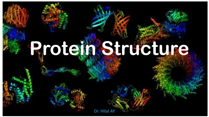
Protein Structure Dr. Hilal AY
What What are are proteins proteins? ?
What What are are proteins proteins? ? Proteins are biological molecules performing a wide variety of functions. Some proteins catalyze a reaction, i.e. they make it go faster than it normally would: those proteins are called enzymes. Other proteins transport molecules throughout the body, others yet provide structural support for cells so they have the right shape, etc.
What are proteins? Proteins are involved in nearly every biological process and their function is very often tightly linked to their three- dimensional structure. Therefore, it is crucial to determine the structure of a protein in order to understand fully how it works inside a cell. Figure 1. Examples of the relationship between protein structure and function. ( a ) Crystal structure of the Lambda-phage repressor (Protein Data Bank (PDB) ID: 3bdn), which binds to its target DNA sequence (red) by making sequence-specific contacts through the grooves in the DNA double- helix; ( b ) Crystal structure of Tsx (PDB ID: 1tlz [5]), a nucleoside transporter protein, that transports nucleosides (red) by creating pores in the membrane through a -barrel motif (shown here in blue); and ( c ) Crystal structure of keratin (PDB ID: 3tnu), a fibrous structural protein whose toughness can be attributed to the helical coiled-coil structure it adopts in its fibers (Burger, V. M., Gurry, T., & Stultz, C. M. (2014). Intrinsically disordered proteins: where computation meets experiment. Polymers, 6(10), 2684-2719.)
What are proteins? The many functions of proteins are reflected by the wide variety of 3D structures they adopt. However, all proteins are made of the same constituents: amino acids.
What are amino acids? Proteins are polymers: similar molecules (called monomers) are repeated many times to form a chain (the polymer). The monomers making up proteins are amino acids.
What are amino acids? Amino acids (except glycine) contain a central chiral carbon, i.e. a carbon atom covalently linked to four different groups of atoms, often called carbon (C ). This central chiral atom is linked to an amino group and a carboxylic acid group, thus the term amino acid. It is also attached to a hydrogen atom and a side chain (sometime called R group). An amino acid s side chain is what sets it apart from the other amino acids and is often responsible for the special chemical and biological properties of the amino acid.
What are amino acids? In water at pH 7 (which is close to physiological conditions inside cells), the amino group is protonated and positively charged, while the carboxylic acid group is deprotonated and negatively charged. An amino acid in water at pH 7 has a positively charged amino group and a negatively charged acid group, but is neutral overall (as the two charges cancel out). Such compounds are called zwitterions.
There are twenty standard side chains (R groups), thus there are twenty standard amino acids.
What are amino acids? Plants can make all of them, whereas animals can only synthesize some of them. The others must be acquired through the diet and are called essential amino acids.
What are the 20 standard amino acids? What are the 20 standard amino acids? There are twenty standard amino acids, which can be grouped according to their properties. Each amino acid can be identified by its name (e.g. Alanine), its three-letter code (e.g. Ala) and its one-letter code (e.g. A).
What are the 20 standard amino acids? What are the 20 standard amino acids?
What are the 20 standard amino acids? What are the 20 standard amino acids?
What are the 20 standard amino acids? What are the 20 standard amino acids?
What are the 20 standard amino acids? What are the 20 standard amino acids?
At pH 7, aspartic acid and glutamic acid lose the proton on their side chain carboxylic acid, making them negatively charged.
Lysine and arginine, on the other hand, gain a proton attached to a nitrogen atom in their side chain, making them positively charged. At pH 7, a small proportion of histidine molecules will have an extra hydrogen attached to one of the nitrogen atoms in the side chain, making the side chain positively charged.
Some amino acids may belong to several categories, depending on the conditions in which they find themselves.
What is the peptide bond? To make a protein, these amino acids are joined together in a polypeptide chain through the formation of a peptide bond.
What is the peptide bond? What is the peptide bond?
What is the peptide bond? What is the peptide bond?
What is the peptide bond? The ( , ) angles of amino acids in a polypeptide chain or protein are restricted, largely because of steric interactions (glycine is an exception).
Primary Structure of Proteins The primary structure of a protein is the linear sequence of the side chains that are connected to the protein backbone.
Secondary Structure of Proteins Primary structure leads to secondary structure; the local conformation of the polypeptide chain or the spatial relationship of amino acid residues that are close together in the primary sequence. In globular proteins, the three basic units of secondary structure are the helix, the strand and turns. All other structures represent variations on one of these basic themes.
Tertiary Structure of Proteins The tertiary structure of a protein refers to the bending and folding of the protein into a specific three-dimensional shape.
Tertiary Structure of Proteins These structures result from four types of interactions between the R side chains of the amino acids residues: 1. Disulfide bridges 2. Salt bridges 3. Hydrogen bonds 4. Hydrophobic interactions
Quaternary Structure of Proteins When two or more polypeptide chains are held together by disulfide bridges, salt bridges, hydrogen bond, or hydrophobic interactions, forming a larger protein complex. Each of the polypeptide subunits has its own primary, secondary, and tertiary structure. The arrangement of the subunits to form a larger protein is the quaternary structure of the protein.
