Pulmonary Embolism and Its Risk Factors
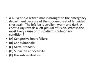
Pulmonary embolism is an obstructive lesion in the pulmonary vascular tree, commonly caused by a thrombus traveling from a distant site. It presents with symptoms like dyspnea and chest pain. Risk factors include recent surgery, immobility, pregnancy, and clotting disorders. Correct diagnosis is challenging, and it can be fatal if untreated. Learn more about this critical condition and its implications.

Download Presentation

Please find below an Image/Link to download the presentation.
The content on the website is provided AS IS for your information and personal use only. It may not be sold, licensed, or shared on other websites without obtaining consent from the author.If you encounter any issues during the download, it is possible that the publisher has removed the file from their server.
You are allowed to download the files provided on this website for personal or commercial use, subject to the condition that they are used lawfully. All files are the property of their respective owners.
The content on the website is provided AS IS for your information and personal use only. It may not be sold, licensed, or shared on other websites without obtaining consent from the author.
E N D
Presentation Transcript
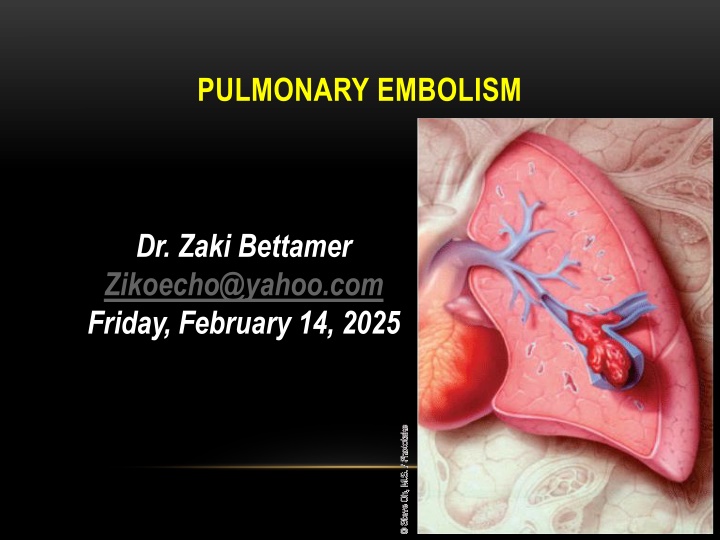
PULMONARY EMBOLISM Dr. Zaki Bettamer Zikoecho@yahoo.com Friday, February 14, 2025
67 years Old male presented with acute dyspnea, Pleuritic Right side chest pain and hemoptysis , with history of recent pelvic fracture post RTA one week back. On examination : Left leg swelling and tender, BP: 90/60, pulse : 140 beat / minute, regular. Chest examination: normal The commonest arrhythmia expected in this patient
DEFINITION: It is an obstructive lesion in the pulmonary vascular tree. It is usually caused by a insoluble thrombus that has travelled from a distant site. A blood clot that forms and remain in a vein is called thrombus, a clot that travels to another part is called embolus.
INCIDENCE: The majority (80%) of pulmonary emboli arise from the propagation of lower limb DVT The annual incidence is about 60-70 per 100 000 population. Rare causes include septic emboli (from endocarditis affecting the tricuspid or pulmonary valves), tumour (especially choriocarcinoma), fat, air, amniotic fluid and placenta. It is a common mode of death in patients with cancer, stroke and pregnancy.
Correct diagnosis is made only in 10-30 %. 60 % of autopsies showed some evidence of post PE. 15 % of postoperative deaths are related to PE or its sequels. PE is a commonest cause of maternal death in the UK.
PREDISPOSING FACTORS Recent surgery especially pelvic, large joint and extensive abdominal . Following pelvic and leg fractures. Pregnancy and child birth. Old age: > 70 years. Smoking . Inherited clotting disorders: a. Hypercoagulable state. b. Platelets abnormalities. c. Protein S & C deficiency.
Long periods of immobility: a. Prolonged hospitalization. b. Long plane or car trips. c. Prolonged bed rest. Severe debilitating illness: a. Severe injures. b. Cardiac diseases. c. Malignancies.
Increased estrogen level: a. Contraceptive pills. b. Hormonal therapy. Increased levels of clotting factors: a. pancreatic, lung and ovarian malignancies (increased procoagulant). b. Female hormones.
Family history. Pace makers or venous catheterization either central or peripheral. Being overweight: There is a link between the formation of clots formation to leptin ( hormone produced by fat cells). Previous proved history of venous thromboembolism.
Cardiac problems: a. Congenital. b. Congestive. c. Hypertensive. Others: a. Nephrotic syndrome. b. Thrombotic and myeloproliferative disorders. c. Inflammatory bowel diseases.
SOURCES AND ORIGIN OF PE The source can be indentified in only 50 70 % of cases. 70 90 % of the identified cases, clots come from IVC. 10 20 % of the identified cases, clotscome from SVC.
PATHOLOGICAL LEADING CAUSES Deep vein thrombosis. Air embolism. Amniotic fluid embolism. Fat embolism. Rare : Tumor emboli & Bacterial clumps.
PROGRESSION OF PULMONARY EMBOLISM. The course of events depend on the size of the clot causing PE. A. Small to medium sized PE. Cut off the circulation to a part of the lung. It heals with fibrous scar. Can travel more distally infracting the lung.
B. Massive pulmonary embolism. Embolize the main pulmonary artery Blocking off almost blood flow to lungs. The emboli may happen suddenly causing Massive PE or gradual causing recurrent PE state. Lodge at the bifurcation of the main pulmonary artery causing hemodynamic collapse or death.
PROBABLE OUTCOME OF PE. 10 % of clinically presented PE are fatal. 30 % of patients with PE will suffer a second attack. 70 % will had PE complications.
DIFFERENTIAL DIAGNOSIS OF PE Myocardial infarction. Pneumonia. Congestive heart failure. Asthma. COPD. Intrathoracic neoplasm. Rib fractures. Pneumothorax. Musculoskeletal pains.
PRESENTATIONS OF PE. ACCORDING TO THE SIZE OF VENOUS EMBOLI: Acute Chronic Large sized. Small sized. Massive PE. Pulmonary infarction with episodes of pleuritic chest pain And haemoptysis. It involves > 50 % of the main PA Sudden death.
POSTOPERATIVE PRESENTATIONS. Classical time: 2nd postoperative week ! Sudden death Cyanosis Dysponea Tackyponea Cough Hemoptysis Chest pains Low EtCO2
SYMPTOMS MORE CLEAR IN PATIENT WITH PRE- EXISTING LUNG DISEASES. POSSIBLE PRESENTATIONS: Apprehension Completely normal. Chest pain Pleuritic pain Dysponea Cough Hemoptysis Diaphoresis
Syncope Tachyponea Leg pain Wheezing Angina pain. AF Coma Pul.infection Sweating Rales Tachycardia Fever > 37.8 Phlebitis Cyanosis Leg swelling
CHRONIC EFFECTS OF PE 1. Increased PVR. 2. Decrease gasexchange. 3. Alveolar hyperventilation. 4. Increased AWP. 5. Decreased lung compliance. 6. Right side ventricular dysfunction & failure.
SCREENING AND DIAGNOSIS. PE is difficult to diagnose especially with underlying lung or cardiac diseases. More than one test should be done to increase the efficacy of correct diagnosis.
1ST LINE OF INVESTIGATIONS. ECG. Nonspecific changes are common. Sinus tachycardia is the commonest among changes. Possible changes: AF, RBBB, T wave inversion & P pulmonale. S1 , Q3, T3
ECG Signs of right ventricular strain (S1, Q3, T3) S wave in lead 1 Q wave in III inverted T in III
CHEST X RAY. POSSIBLE FINDINGS Elevated diaphragm. normal. Prominent PA. Atelectasis or density. Cardiomegally. Pleural effusion. Pulmonary edema. Pleural opacity.
Wedge shaped density. Pleural effusion
ABGS May be normal ! Hypoxia. Respiratory hypocarbia. Increased A-a gradient. * Gradient > 15-20 is considered abnormal.
D DIMER TEST. D dimer are released as a result of fibrinolysis & indicate the presence of intravascular thrombus. It reflects the breakdown of plasmin. Useful, to exclude the PE. Has > 99% sensitivity in acute PE or DVT. .
Negative D dimer Test. Exclude PE in Patient with low pre-test Probability. Helpful in patient with a high clinical Probability. Positive D dimer test. Needs further investigations to confirm the diagnosis Less useful in patients with longer hospitalization. False positive can result from the clot formation at venepuncture sites.
FALSE POSITIVE D DIMER TEST Malignant Patient. Recent Surgery. Chronic Inflammatory Process Sepsis Venepuncture Due to clot formation
PULMONARY ANGIOGRAPHY Shows pulmonary vascular obstruction Gold standard of diagnosis of PE, Thrombolytic therapy can be given through the catheter afterwards.
COMPUTED TOMOGRAPHIC PULMONARY ANGIOGRAM Has sensitivity > 90 % Dilatation of pulmonary vessels proximal to the embolus with collapse of distal vessels.
ISOTOPE LUNG SCAN. ( VENTILATION/PERFUSION SCAN) Use of radioactive tracers to study air flow( ventilation ) and blood flow (perfusion).
2ND LINE OF INVESTIGATIONS. Leg ultrasound scan doppler for detection of DVT. Around 70 % of patient proved PE will have a proximal DVT. . Echocardiogram. Diagnostic only in the massive PE. Of prognostic information's. The findings may be: * RV overload & dysfunction. * Dilatation of IVC. * Dilated pulmonary arties. * Disturbed flow velocity. * Direct visualization of clots in the prox. PA, RV, or RA.
SPIRAL HELICAL COMPUTERIZED TOMOGRAPHY. Three dimensional series of very thin x ray beams using contrast dye.
LUNG ULTRASOUND. 1.It is noninvasive sonar. 2. Uses high frequency sound waves to check for blood clots. 3. It is cheaper than V/Q scan Venography. Invasive procedure to reveal clot blockage at any point of the vein course using contrast.
MAGNETIC RESONANCE ( MRI ). Expensive. Reserved for pregnant woman and renal patient. TEE. Intraoperative diagnosis of PE.
C. INTRAOPERATIVE PROPHYLAXIS Decrease venousstasis: 1. Raising legs. 2. Avoid leg trauma. 3. Use of pneumatic leg compression. 4. Use of electrical calf muscle stimulation.
WHEN PULMONARY EMBOLISM IS LIFE THREATENING ? Massive PE. Worsening the cardiopulmonary underlying diseases. Treatment plan are not effective.
TREATMENT OVERVIEW OF PE 1. Preventive strategies plan: A. Preventive steps in the hospital: 1. Correction of the risk factors. 2. Adequate hydration. 3. Graduated compression stockings. 4. Use of pneumatic compression. 5. Early physical activity.
6. Anticoagulant therapy. a. Heparin:5000 iu, 8-12 h 1 , 2h before operation. b. Ultra low dose heparin: 1 unit.kg.h 1 infusion. c. Adjusted heparin dose: start 3500 iu subcut. Every 8 h 1 to keep ATT 1.5 X normal. d. Low molecular heparin:one dose, 12h 1 subcut.untill patient ambulate.
B. PREVENTIVE STEPS WHILE TRAVELLING. Take a walk. KtoKkid_walking_sm_wht Exercise while you set. Wear support stockings. Drink plenty of fluids. Ask for medical advise: * If you had DVT before. * If the travelling time is more than 6 hours. * If you at any risk.
II. ACTIVE STRATEGIES PLANS. A. Treatment of DVT: 1. Heparin. * 5000 IU bolus, followed by 40000 IU over 24 h for 7 days. * adjust the dose: APTT 1.5-2 2. Oral anticoagulants: * Warfarin for 3 6 month. * Keep PT 1.2-1.5 ( INR : 2 3 )
B. Thrombolytic therapy. indicated for acute massive PE causing collapse or arrest. 1. Streptokinase: 250000 U IV over 30 min, then 100000 U h 1 for 24 hours. 2. Tissue type plasmogen activator 100 mg IV over 2 hours. 3. Urokinase: 4400 IU Kg 1 over 10 min. followed by 4400 IU Kg 1.h 1 for 12 -24 hours.
Actilyse Alteplase * Action: It is an enzyme that increase plasmin that break down clots. Recommendations a. Should be given as soon as possible. b. Given as infusion over 2-3 hours. c. 10 mg bolus over one or two minutes followed by 90 mg.
C. Surgical pulmonary embolectomy. * Under CP bypass. * Postoperative & operative mortality is 20 50 % * Long term survival is 71 % after 5 years. * Indicated in acute massive PE with life D. I.V.C umbrella filter: * Placed percutaneously under local analgesia. * Aim: to avoid recurrence of more emboli.
D. I.V.C umbrella filter: * Indications: a. Contraindication to anticoagulant. b. Failure of anticoagulant therapy. c. Chronic recurrent PE d. Emergency treatment following massive PE.


.")