Rabies: Symptoms, Transmission, and Pathogenesis
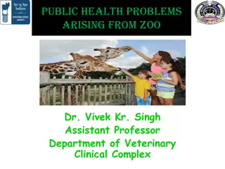
Rabies is a deadly virus transmitted through the saliva of infected animals, with common carriers being bats, coyotes, foxes, raccoons, and skunks. Symptoms include fever, headache, nausea, and hydrophobia. The rabies virus, a member of the Rhabdoviridae family, travels to the central nervous system, leading to fatal encephalitis. Prevention through rabies vaccinations is crucial, as treatment after symptoms appear is nearly always unsuccessful.

Download Presentation

Please find below an Image/Link to download the presentation.
The content on the website is provided AS IS for your information and personal use only. It may not be sold, licensed, or shared on other websites without obtaining consent from the author. If you encounter any issues during the download, it is possible that the publisher has removed the file from their server.
You are allowed to download the files provided on this website for personal or commercial use, subject to the condition that they are used lawfully. All files are the property of their respective owners.
The content on the website is provided AS IS for your information and personal use only. It may not be sold, licensed, or shared on other websites without obtaining consent from the author.
E N D
Presentation Transcript
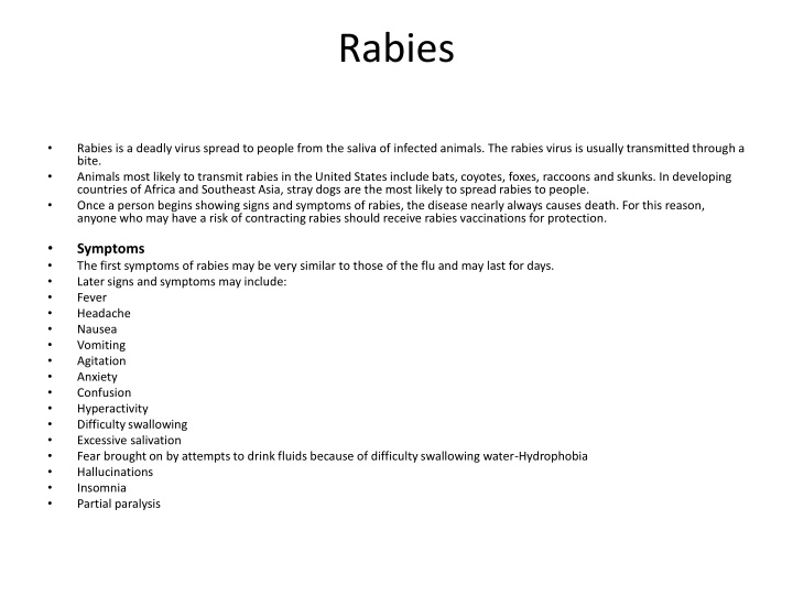
Rabies Rabies is a deadly virus spread to people from the saliva of infected animals. The rabies virus is usually transmitted through a bite. Animals most likely to transmit rabies in the United States include bats, coyotes, foxes, raccoons and skunks. In developing countries of Africa and Southeast Asia, stray dogs are the most likely to spread rabies to people. Once a person begins showing signs and symptoms of rabies, the disease nearly always causes death. For this reason, anyone who may have a risk of contracting rabies should receive rabies vaccinations for protection. Symptoms The first symptoms of rabies may be very similar to those of the flu and may last for days. Later signs and symptoms may include: Fever Headache Nausea Vomiting Agitation Anxiety Confusion Hyperactivity Difficulty swallowing Excessive salivation Fear brought on by attempts to drink fluids because of difficulty swallowing water-Hydrophobia Hallucinations Insomnia Partial paralysis
Rhabdoviruses are simple viruses encoding only five proteins and appearing as bullet-shaped enveloped virions with a diameter of 50 to 95 nm and length of 130 to 380 nm Within the envelope, the helical nucleocapsid is coiled symmetrically into a cylindrical structure, giving it the appearance of striations . The nucleocapsid is composed of one molecule of single-stranded, negative sense ribonucleic acid (RNA) of approximately 12,000 bases and the nucleoprotein (N), large (L), and nonstructural (NS) proteins. The L and NS proteins constitute the RNAdependent RNA polymerase. The N protein is the major structural protein of the virus. It protects the RNA from ribonuclease digestion and maintains the RNA in a configuration acceptable for transcription. The matrix (M) protein lies between the envelope and the nucleocapsid
Pathogenesis Rabies infection usually results from the bite of a rabid animal. Rabies infection of the animal causes secretion of the virus in the animal s saliva and promotes aggressive behavior ( mad dog), which in turn promotes transmission of the virus. The virus can also be transmitted through inhalation of aerosolized virus (as may be found in bat caves), in transplanted infected tissue (e.g., cornea), and by inoculation through intact mucosal membranes. The virus replicates quietly at the site for days to months (Figure) before progressing to the central nervous system (CNS). Rabies virus travels by retrograde axoplasmic transport to the dorsal root ganglia and the spinal cord. Once the virus gains access to the spinal cord, the brain becomes rapidly infected. The affected areas are the hippocampus, brainstem, ganglionic cells of the pontine nuclei, and Purkinje cells of the cerebellum. The virus then disseminates from the CNS via afferent neurons to highly innervated sites such as the skin of the head and neck, salivary glands, retina, cornea, nasal mucosa, adrenal medulla, renal parenchyma, and pancreatic acinar cells. After the virus invades the brain and spinal cord, encephalitis develops and neurons degenerate. Despite extensive CNS involvement and impairment of CNS function, little histopathologic change can be observed in the affected tissue, other than the presence of Negri bodies Rabies is fatal once clinical disease is apparent. The length of the incubation period is determined by the (1) concentration of the virus in the inoculum, (2) proximity of the wound to the brain, (3) severity of the wound, (4) host s age, and (5) host s immune status. Cell-mediated immunity appears to play little or no role in protection against rabies virus infection. Antibody can block the spread of virus to the CNS and brain if administered or generated by vaccination during the incubation period. The incubation period is usually long enough to allow generation of a therapeutic protective antibody response after active immunization with the killed rabies vaccine.
Laboratory Diagnosis The occurrence of neurologic symptoms in a person who has been bitten by an animal generally establishes the diagnosis of rabies. Laboratory tests are usually performed to confirm the diagnosis and determine whether a suspected individual or animal is rabid (postmortem). Antigen detection using direct immunofluorescence genome detection using reverse transcriptase polymerase chain reaction (RT-PCR) are relatively quick and sensitive assays that are the preferred methods for diagnosing rabies. Samples of saliva are easy to test, but serum, spinal fluid, skin biopsy material from the nape of the neck, brain biopsy or autopsy material, and impression smears of corneal epithelial cells can also be examined. Infected cells will have intracytoplasmic inclusions consisting of aggregates of viral nucleocapsids (Negri bodies) in affected neurons. . Rabies antibody titers in serum and cerebrospinal fluid are usually measured by enzyme-linked immunosorbent assay (ELISA). Antibody usually is not detectable until late in the disease.
Treatment and Prophylaxis Clinical rabies is almost always fatal unless treated with post rabies immunization. Postexposure prophylaxis is the only hope for preventing overt clinical illness in the affected person. The wound should be washed immediately with soap and water or another substance that inactivates the virus. Antirabies immunoglobulin is injected near the wound. Subsequently, four immunizations with rabies vaccine are administered within 2 weeks(The rabies vaccine is a killed-virus vaccine prepared through chemical inactivation of rabies infected tissue culture human diploid cells (HDCV) or chick embryo cells. ) Passive immunization with human rabies immunoglobulin(HRIG) provides antibody until the patient produces antibody in response to the vaccine