Radiology Anatomy of Upper Limb: Practical Insights and Imaging
Explore detailed radiology images of the upper limb, including the shoulder, elbow, and hand. Learn about different structures such as the acromion, humerus, and deltoid muscle through X-rays, MRIs, and CT scans. Understand common conditions like fractures and muscular anatomy for a comprehensive study of upper limb radiology.

Download Presentation

Please find below an Image/Link to download the presentation.
The content on the website is provided AS IS for your information and personal use only. It may not be sold, licensed, or shared on other websites without obtaining consent from the author. If you encounter any issues during the download, it is possible that the publisher has removed the file from their server.
You are allowed to download the files provided on this website for personal or commercial use, subject to the condition that they are used lawfully. All files are the property of their respective owners.
The content on the website is provided AS IS for your information and personal use only. It may not be sold, licensed, or shared on other websites without obtaining consent from the author.
E N D
Presentation Transcript
RADIOLOGY ANATOMY OF UPPER LIMB Practical (2) Radiology
acromion Clavicale Greater teberosity coracoid Lesser teberosity scapula glenoid
acromion Y view Humeral head spine coraocid scapula humors shaft
Humerus fracture
acromion clavicle Supraspinatus muscle Humeral head Deltoid muscle Glenoid cavity Subscapularis muscle Teres minor muscle
1-Biceps Muscle. 2-Brachialis Muscle. 3-Brachial Artery. 4-Humerus. 5-Triceps Muscle
Olecranon fossa medial epicondyle Lateral epicondyle capitulum trochlea Radial head ulna Radial teberosity
humerus Coroniod process Radial teberosity Olecranon fossa Radial head olecranon ulna
CHILD ADULT
Hock of hamate Ulnar styloid process
Distal phalanx Distal interphalanx joint Middle phalanx Proximal interphalanx joint Proximal phalanx Metacarpophalangeal joint
CHILD ADULT
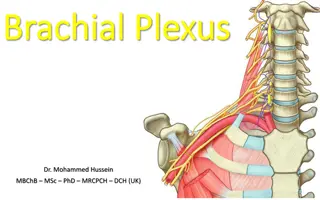
1-Vertebral Artery. 2-Axillary Artery. 3-Internal Thoracic Artery. 4-Posterior Humeral Circumflex Artery. 5-Circumflex Scapular Artery. 6-Subscapular Artery 7-brachial artery
1-Radial Artery. 2-Ulnar Artery. 3-Deep Palmar Arch. 4-Common Palmar Digital Artery. 5-Proper Palmar Digital Artery
RT. UPPER EXTREMITY ANGIOGRAM RADIAL ARTERY ELBOW JOINT INTEROSSEOUS ARTERY ULNAR ARTERY BRACHIAL ARTERY RT
RT. UPPER EXTREMITY ANGIOGRAM PALMAR ARCH RADIAL ARTERY ULNAR ARTERY RT