Radiology and the Kidney: Clinical Cases and Diagnostic Techniques
Explore the diagnostic approach to kidney-related cases through radiology imaging techniques, including diuretic renal scans and obstructive uropathy evaluation. Learn about common indications and studies for assessing renal conditions in patients.

Download Presentation

Please find below an Image/Link to download the presentation.
The content on the website is provided AS IS for your information and personal use only. It may not be sold, licensed, or shared on other websites without obtaining consent from the author. If you encounter any issues during the download, it is possible that the publisher has removed the file from their server.
You are allowed to download the files provided on this website for personal or commercial use, subject to the condition that they are used lawfully. All files are the property of their respective owners.
The content on the website is provided AS IS for your information and personal use only. It may not be sold, licensed, or shared on other websites without obtaining consent from the author.
E N D
Presentation Transcript
Radiology and the Kidney Brian Cronin MD, FASN, FACP March 13 2025
Disclosure Some questions modified from ASN KSAP NephSAP
Case 1 57 yo female presents with right sided abdominal pain. PMH: Solitary kidney s/p left nephrectomy at age of 11 for dysfunctional kidney incidentally found on imaging done for trauma. PE: BP 131/65 HR 67 NAD, abdomen soft, nontender no palpable masses, no edema Cr 1.19 (eGFR 53), baseline 0.91, improved to 0.85 over 12 hrs with IVF UA: SG 1.008, neg prot, 1+ blood, neg leukocyte, 2 RBC, 1 WBC per HPF CT (w/o contrast): Moderate to severe right hydronephrosis with abrupt narrowing of the ureteropelvic junction. What study is best to assess for obstruction?
Diuretic Renal Scan A nuclear medicine renal scan involves imaging with a compound taken up by the kidney that is bound to a radioisotope. There are different compounds used that are bound to the radioisotope technetium: MAG3: 99m dimercaptosuccinic acid Most common compound used for renal scans Rapid extraction by renal tubular cells. Higher extraction rate than DTPA. Secretion is mainly by proximal tubules
Evaluation of Obstructive Uropathy: Diuretic (Furosemide) Renal Scan Renal scanning is often used to assess if hydronephrosis is due to true functional obstruction as opposed to anatomic non- obstructive dilatation. Non obstructive hydronephrosis can be seen in vesicoureteral reflux, congenital malformations, or with a noncompliant bladder. In these cases a diuretic (furosemide) renal scan is performed. The radionuclide (typically bound to MAG3) is injected. Renal excretion of the radiotracer is assessed. Diuretic (furosemide) is administered. If radiotracer excretion increases (normal half life < 9.8 minutes), indicative of non obstructive dilatation. If radiotracer excretion unaffected or minimally effective (prolonged half life > 20 minutes) consistent with obstructive uropathy NSAIDS should not be administered as affect radionuclide excretion
Indications for Renal Scan * Common Uses
Case 2 58 year old male referred for stage 3 CKD. PMH: HTN, DM, hyperlipidemia SH: 10 pack year history of smoking, quit 25 years ago Medications: Lisinopril, dapagliflozin, rosuvastatin, aspirin PE: BP 146/84 HR 72, NAD. No edema Labs: C4 1.54 (eGFR 52), UA: trace protein; neg blood; neg leukocyte, UACR 240 mg/g US: Normal size and echogenicity. 3 cm cyst with complex features in the right upper pole What is the best study to assess the cyst?
CT Urogram CT urogram: 3 phase CT with contrast Non contrast phase Nephrogenic phase Excretory phase Test of choice for urologic evaluation of hematuria Contrast required to rule out renal malignancy Benign lesions do not enhance Renal cell carcinomas enhance Oncocytomas and lipid poor angiomyolipomas (benign neoplasms) also enhance
Case 3 29 year old female with family history of ADPKD presents for routine physical. She asks if there is testing to determine if she has PKD. ROS: Negative for hematuria or flank pain PE: BP 108/74 HR 72 remainder normal Cr 0.72, eGFR 116, UA: negative occult blood, protein, leukocyte Can ultrasound rule out ADPKD?
ADPKD Age # of Cysts PPV: Sensitivity: 15-29 > 3 total PPV: 100% Sensitivity: 69.5 - 94.3% 30-39 > 3 total PPV: 100% Sensitivity: 94.9 - 96.6% 40-59 > 2 in each kidney PPV: 100% Sensitivity: 88.8 - 92.6%
Case 4 70 yo male with HTN, CAD, hyperlipidemia and recent right total knee replacement presents with acute onset dyspnea and pleuritic chest pain. . PE: BP 158/74; HR 108; 88% on RA mildly dyspneic, no murmurs, lungs CTA, neg edema CXR: Mild atelectasis, cardiomegaly Cr 1.84, eGFR 39 (at baseline) Do you order CTA chest with contrast?
Contrast Nephropathy Contrast Associated Acute Kidney Injury (CA-AKI): AKI that occurs after IV contrast. Contrast Induced Acute Kidney Injury (CI-AKI): AKI that occurs because of contrast.
Contrast Nephropathy Everything in medicine is a risk benefit ratio. The risk is either high, low, or theoretically possible. The benefit is either high, low, or theoretically possible. If the benefit of contrast is extremely high, it may make sense to give the contrast even with CKD 4 or 5 (eGFR < 30) or AKI (see below). If the benefit of contrast is very low, it may make sense to not give contrast for lesser degrees of CKD (ie. CKD stage 3). The risk assessment is for CKD not AKI. In AKI the eGFR does not accurately reflect the true level of kidney function because it is based on the creatinine (which lags behind the true eGFR). The ACR Manual On Contrast Media says: In fact, since each contrast medium administration always implies a risk-benefit analysis for the patient, contrast medium administration for all patients should always be taken in the clinical context, considering all risks, benefits and alternatives patients with AKI are particularly susceptible to nephrotoxin exposure and therefore it is probably prudent to avoid intravascular iodinated contrast medium in these patients when possible.
Contrast Nephropathy ACR Manual On Contrast Media recommends prophylaxis with Normal Saline for: 1. Patients with AKI. 2. CKD with eGFR < 30 (not dialysis). 3. Individual high risk cases with eGFR 30-44 at the discretion of the physician (borderline GFR, recent AKI, numerous risk factors for AKI) being the considerations
Case 5 61 yo female with type 2 DM, HTN, CKD; has several weeks of diplopia and headache. Contrast enhanced brain MRI ordered by neurology. Cr 2.23, eGFR 24 What do you recommend? MRI without contrast CT with contrast MRI with group 2 contrast MRI with contrast followed by dialysis
MRI Contrast MRI contrast is a substance called gadolinium. This differs from iodinated contrast. Gadolinium is a rare earth metal that has strong paramagnetic properties, making it ideal for MR imaging. Free gadolinium is toxic, therefore it must be chelated to an organic ligand to encapsulate the ion and reduce toxicity. These organic ligands can be either linear or macrocyclic, ionic or nonionic.
Is Gadolinium Nephrotoxic? Gadolinium has been shown to affect glomerular filtration rate (GFR), however these effects are typically minimal, transient and not clinically significant. Therefore, gadolinium is not considered to be nephrotoxic. For this reason MR angiography (in lieu of CT angiography) with gadolinium became the imaging of choice for patients with CKD, ESRD or AKI.
Nephrogenic Systemic Fibrosis Nephrogenic Systemic Fibrosis (NSF) was observed in patients with advanced CKD or ESRD. It is a scleroderma like condition often resulting in debilitating and even fatal tissue and organ fibrosis. It manifests as: Cutaneous thickening or indurated, discolored skin with plaques, papules and/or nodules. Affects trunk and limbs, in a bilateral, symmetric manner. Starts distal and progresses proximal. Spares face. Sclerodactyly and joint contractures may occur. Multiorgan involvement with internal organ fibrosis may occur.
Nephrogenic Systemic Fibrosis It was observed that nephrogenic systemic fibrosis (NSF) was associated with linear, but not macrocyclic gadolinium agents. Macrocyclic ligands are more stable than linear ones, therefore associated with a lower risk of free gadolinium deposition. However, macrocyclic agents have a higher incidence of allergic reactions. The different gadolinium agents were then classified by the American College of Radiology based on their risk of developing NSF. Group I Greatest number of associated NSF cases Group II Few, if any associated NSF cases Group III Few, if any associated NSF cases, but limited data
Gadolinium in CKD Regarding the risk of NSF the general consensus is: Any type of gadolinium is safe with an eGFR> 30 Group II agents should be used with eGFR < 30 or ESRD Gadolinium is cleared by dialysis, therefore some suggest scheduling dialysis in an ESRD patient after administration if possible. Most do not recommend extra dialysis or performing dialysis in a patient with AKI or CKD stage 4 or 5 not on dialysis.
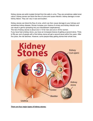
Case 6 50 yo male presents with sudden and severe right flank pain. No associated fever or gross hematuria. He recalls spontaneously passing a kidney stone 10 years ago. He eats a high protein diet and does a lot of exercise in the gym. Serum creatinine normal UA: trace protein, 20-50 RBC/hpf What study do you order? Abdominal X-ray (KUB) Renal Ultrasound CT with Contrast CT without contrast
KUB X Ray Only Detects Radiopaque Stones Radiopaque Radiolucent Calcium oxalate Uric acid Calcium phosphate (poorly) Drug Stones Struvite (poorly) Xanthine Cystine (poorly) 2,8 dihydroxyadenine
KUB X Ray Low Sensitivity and Specificity Uses: Surveillance Post Urologic Intervention
Ultrasound Low Sensitivity to Detect Stones (50%) Very Low Sensitivity to Detect Ureteral Stones High Sensitivity to Detect Hydronephrosis (90%) No Radiation. Can be Repeated Frequently
CT Unenhanced CT: Gold Standard Sensitivity: 97% Specificity: 98-99% Radiation Exposure
Questions? BC Nephro bcnephro.com Brian Cronin -YouTube instagram.com/bcnephro/?hl=en


")