Explore the case of a 50-year-old male with hematuria who underwent sonography, encountering an echogenic band obscuring part of the urinary bladder image due to range ambiguity artifact, caused by high pulse repetition frequency and large field depth. Learn about the formation of B-mode ultrasound images, the impact of multiple focal zones, and how to recognize and correct this artifact clinically.
Please find below an Image/Link to download the presentation.
The content on the website is provided AS IS for your information and personal use only. It may not be sold, licensed, or shared on other websites without obtaining consent from the author. If you encounter any issues during the download, it is possible that the publisher has removed the file from their server.
You are allowed to download the files provided on this website for personal or commercial use, subject to the condition that they are used lawfully. All files are the property of their respective owners.
The content on the website is provided AS IS for your information and personal use only. It may not be sold, licensed, or shared on other websites without obtaining consent from the author.
E N D
Presentation Transcript
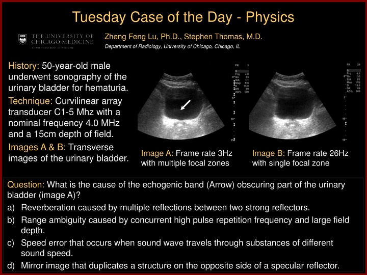
Tuesday Case of the Day - Physics Zheng Feng Lu, Ph.D., Stephen Thomas, M.D. Department of Radiology, University of Chicago, Chicago, IL History: 50-year-old male underwent sonography of the urinary bladder for hematuria. Technique: Curvilinear array transducer C1-5 Mhz with a nominal frequency 4.0 MHz and a 15cm depth of field. Images A & B: Transverse images of the urinary bladder. Image A: Frame rate 3Hz with multiple focal zones Image B: Frame rate 26Hz with single focal zone Question: What is the cause of the echogenic band (Arrow) obscuring part of the urinary bladder (image A)? a) Reverberation caused by multiple reflections between two strong reflectors. b) Range ambiguity caused by concurrent high pulse repetition frequency and large field depth. c) Speed error that occurs when sound wave travels through substances of different sound speed. d) Mirror image that duplicates a structure on the opposite side of a specular reflector.
Discussion A B-mode ultrasound image is formed by emitting a pulse and collecting the resultant echo signal along the insonified path. After each transmit pulse, sufficient time must be allocated for the echoes to return using the following range equation: D = ct/2 Where D is the depth, t is the delay time between pulse emission and echo reception and c is the sound propagation speed. Echoes from a deeper depth takes a longer time to return. When echoes from deep structure from the previous transmit pulses are received simultaneously as the echoes from shallow structure insonified by the present pulse, the range ambiguity artifact occurs. This typically happens when multiple focal zones are set for a large field-of-view because multiple pulses are needed along the same beam line while each pulse can only focus at one particular depth. Consequently, very high pulse repetition frequency is typically employed for multiple focal zone settings, which causes the range ambiguity artifact especially when the field-of-view is large. This may become more obvious when the object has low attenuation such as a urinary bladder. By reducing the number of focal zones to one, the pulse repetition frequency is reduced; thus the range ambiguity artifact is removed. Clinically, range ambiguity artifact is important to recognize and correct as it can obscure important features such as tissue planes and organ pathology.
References RT O Brien, JA Zagzebski and FA Delaney, Ultrasound corner: range ambiguity artifact , Veterinary Radiology & Ultrasound, Vol. 42, No. 6, 2001, pp 542 545. A Goldstein, Range ambiguities in real-time ultrasound , J Clin Ultrasound, Vol 9, No. 2, 1981, pp83-90.


,")

