Referral Mechanisms for Victims of Domestic Violence in Tajikistan
Lack of access to legal and psychological services in rural areas hinders women's ability to protect their rights in Tajikistan. Gender stereotypes and the normalization of domestic violence pose significant challenges. However, NGOs like Najoti Kudakon have made key achievements in lobbying for laws and providing support services for victims, including establishing women support groups and shelters. Despite prevailing cultural beliefs, efforts are being made to address and prevent domestic violence in the country.

Download Presentation

Please find below an Image/Link to download the presentation.
The content on the website is provided AS IS for your information and personal use only. It may not be sold, licensed, or shared on other websites without obtaining consent from the author.If you encounter any issues during the download, it is possible that the publisher has removed the file from their server.
You are allowed to download the files provided on this website for personal or commercial use, subject to the condition that they are used lawfully. All files are the property of their respective owners.
The content on the website is provided AS IS for your information and personal use only. It may not be sold, licensed, or shared on other websites without obtaining consent from the author.
E N D
Presentation Transcript
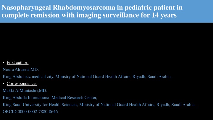
Nasopharyngeal Rhabdomyosarcoma in pediatric patient in complete remission with imaging surveillance for 14 years First author: NouraAlraeesi,MD. King Abdulaziz medical city. Ministry of National Guard Health Affairs, Riyadh, Saudi Arabia. Correspondence: MakkiAlMuntashri,MD. King Abdulla International Medical Research Center, King Saud University for Health Sciences, Ministry of National Guard Health Affairs, Riyadh, Saudi Arabia. ORCID:0000-0002-7880-8646
CASE SYNOPSIS History 18 year old male known case of nasopharyngeal Rhabdomyosarcoma, status post excision and chemoradiotherapy. The patient was diagnosed in 2010, when he presented with difficulty breathing. Imaging Chest x-ray: Soft tissue density projecting in the posterior aspect of the nasopharyngeal airway causing narrowing. Non-enhanced CT of the sinuses: A large soft tissue lesion arising from the nasopharynx extending to the nasal cavity and compressing the adenoid tissue and narrowing the nasopharyngeal airway. Enhanced MRI of the neck: Corresponding mass exhibiting low and high-signal intensity on both T1 and T2 weighted images with post- contrast heterogeneous enhancement. There was no obvious infiltration of the skull base or focal bone destruction. No suspicious cervical lymph nodes. MRI exam of the brain: No evidence of parenchymal lesion or abnormal leptomeningeal enhancement. Bone scan: No scintigraphic evidence of distant skeletal metastases. Pan-CT: Negative. Pathology Management Functional endoscopic sinus surgery with complete excision of nasopharyngeal mass. He received chemotherapy for 1 year and currently he is off treatment since 2011. Yearly MRI follow-up showed no evidence of recurrence. The pathology confirmed the diagnosis of Nasopharyngeal Rhabdomyosarcoma, with no evidence of nodal or distant metastasis. Bone marrow biopsy showed no evidence of bony infiltration.
Diagnostic imaging Lateral cervical spine x-ray; Soft tissue density projecting in the posterior aspect of the nasopharyngeal airway, causing nasopharyngeal airway narrowing Plain CT of the paranasal sinuses; axial and sagittal reformates: Soft tissue density projecting in the posterior aspect of the nasopharyngeal airway displacing the parapharyrngeal fat space laterally, inseparable from the nasal choana. The adenoid tissue is compressed by the mass.
Enhanced MRI of the neck; Axial T2 WI: Well-defined mass arising from the nasopharyngeal mucosa, exhibiting high T2 weighted images , displacing the nasopharyngeal airway laterally, with no invasion. Axial T1 WI post-contrast: heterogeneous enhancement of the mass, no features of vascular, perineural or bony invasion. Sagittal T1 WI post-contrast: Compression of the adenoid tissue, with extension to the nasal cavity. No extension to the oral cavity. Severe narrowing of the nasopharyngeal airway.
Literature references 1. Van Rijn RR, Wilde JC, Bras J, et al. Imaging findings in noncraniofacial childhood rhabdomyosarcoma. Pediatr Radiol. 2008;38:617 634. doi: 10.1007/s00247-008-0751-y. 2. Stevens MC, Rey A, Bouvet N, et al. Treatment of nonmetastatic rhabdomyosarcoma in childhood and adolescence: third study of the International Society of Paediatric Oncology SIOP Malignant Mesenchymal Tumor 89. J Clin Oncol. 2005;23:2618 2628. doi: 10.1200/JCO.2005.08.130. 3. Hicks J, Flaitz C. Rhabdomyosarcoma of the head and neck in children. Oral Oncol. 2002;38:450 459. doi: 10.1016/S1368- 8375(01)00105-1. 4. Kim EE, Valenzuela RF, Kumar AJ, et al. Imaging and clinical spectrum of rhabdomyosarcoma in children. Clin Imaging. 2000;24:257 262. doi: 10.1016/S0899-7071(00)00222-9. 5. J N Healy, M F Borg. Paediatric nasopharyngeal rhabdomyosarcoma: A case series and literature review. Journal of medical imaging and radiation oncology. doi:10.1111/j.1754-9485.2010.02187.x.