Regulation of Enzyme Activity
Enzyme activity is regulated through various mechanisms including enzyme quantity regulation, allosteric regulation, induction, repression, and different models of allosteric regulation. Allosteric effectors can alter enzyme-substrate affinity and catalytic activity. Explore the diverse ways enzymes are controlled for optimal function.

Download Presentation

Please find below an Image/Link to download the presentation.
The content on the website is provided AS IS for your information and personal use only. It may not be sold, licensed, or shared on other websites without obtaining consent from the author.If you encounter any issues during the download, it is possible that the publisher has removed the file from their server.
You are allowed to download the files provided on this website for personal or commercial use, subject to the condition that they are used lawfully. All files are the property of their respective owners.
The content on the website is provided AS IS for your information and personal use only. It may not be sold, licensed, or shared on other websites without obtaining consent from the author.
E N D
Presentation Transcript
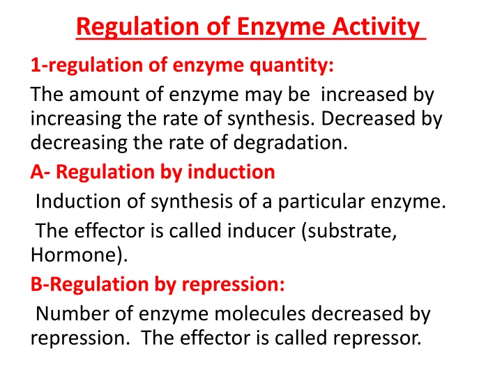
Regulation of Enzyme Activity 1-regulation of enzyme quantity: The amount of enzyme may be increased by increasing the rate of synthesis. Decreased by decreasing the rate of degradation. A- Regulation by induction Induction of synthesis of a particular enzyme. The effector is called inducer (substrate, Hormone). B-Regulation by repression: Number of enzyme molecules decreased by repression. The effector is called repressor.
2- Allosteric Regulation Allosteric Enzymes are regulated by molecules called allosteric effectors (also modifiers) that bind noncovalently at a site ( allosteric site ) other than the active site. These enzymes are composed of multiple subunits, and the regulatory site that binds the effector may be located on a subunit that is not itself catalytic.
The presence of an allosteric effector can alter the affinity of the enzyme for its substrate, or modify the maximal catalytic activity of the enzyme, or both. Effectors that inhibit enzyme activity are termed negative effectors, whereas those that increase enzyme activity are called positive effectors.
Allosteric effectors could be: 1. Homotropic effectors: When the substrate itself serves as an allosteric effector, the effect is said to be homotropic. Most often, an allosteric substrate functions as a positive effector. 2. Heterotropic effectors: The allosteric effector may be different from the substrate. Heterotropic effectors are commonly encountered,for example, the glycolytic enzyme phosphfructokinase allosterically inhibited by citrate, which is not a substrate for the enzyme
MODEL OF ALLOSTERIC REGULATION Two main model have been proposed to describe the mechnastic basis of enzyme allostery: 1-Concerted model : 2-Sequential model:
Concerted ( Symmetry ) Model For an enzyme consists of 2 identical subunits each with one active site. The subunits present in 2 conformations either T ( low affinity ) or R ( high affinity ). An important assumption of the concerted model is that both subunits must be in the same conformational state: the binding of the first substrate molecule to one subunit increases the tendency of all subunits to undergo transition to the high affinity form through an all- or non effect. All subunits being either in the low affinity or high affinity forms. Therefore , in the concerted model, TT , RR conformations are allowed , but TR conformation is not permitted.
2. Sequential Model It postulates that the subunits undergo individual sequential changes in conformation. The binding of the S molecule change the shape of the subunit to which is bound, However, the conformational state of the other subunit is not necessarily altered. Thus, TR conformation is allowed in addition to TT and RR conformations.
3-Feedback inhibition: The end product of metabolic pathway results in allosteric inhibition of the first enzyme in the pathway. Example : For a biosynthetic pathway from A to D Enz 1 Enz 2 Enz 3 A B C D High concentration of D typically inhibit Enzyme 1
4-Zymogens ( Proenzymes) : Are enzymes which are synthesized initially in an inactive form, and then activated at physiologically appropriate time and place. The classical examples are the digestive enzymes: Pepsin , Trypsin and Chymotrypsin which are secreted in an inactive forms . Modes of activation :- Could be Exocatalysis : when a zymogen is activated by another substance. Autocatalysis: when a zymogen is activated by it's already activated precursors.
Examples: 1) HCL exocatalysis Pepsinogen pepsin 2) Pepsin autocatalysis 1) Enterokinase exocatalysis Trypsinogen Trypsin 2) Trypsin exocatalysis Trypsin exocatalysis Chymotrypsinogen Chymotrypsin
5-Covalent Modification :- This is done by reversible covalent insertion of a small group ( ex. phosphate) on certain amino acid residue of an enzyme ( ex. serine, threonine, tyrosine, histidine) resulting in modulation of their activity. Enzymes undergo covalent modification are called Interconvertable enzymes existing in 2 states of activity: one of high catalytic activity and the other of low catalytic activity.
ISOENZYMES Definition: - The multiple forms of an enzyme catalyzing the same reaction are isoenzymes . -Isoenzymes may be present in different tissues of the same organism, in different cell types or subcellular compartments. Besides the source, they also differ from each other with respect to their structure, electrophoretic mobility and immunological properties.
Lactate dehydrogenase (LDH) : There are 5 isozymes of LDH ( 1- 5 ), separated by electrophoresis, all catalyses the same reaction: LDH Lactate + NAD+ pyruvate + NADH + H+ Each isozyme consists of 4 polypeptide chains: H and/or M
LDH ISOZYMES Isozyme No. Subunits Organ Clinical Significance 1 HHHH Heart Myocardial Infarction 2 HHHM Heart, Kidney RBCs Myocardial Infarction Renal cortex infarction, Haemolytic anaemia, 3 HHMM Widely distributed Little significance 4 HMMM Widely distributed Little significance 5 MMMM Liver, skeletal muscle Acute hepatitis, Acute muscle injury, Muscle diseases
Creatine Phosphokinase (CPK) ( Creatine Kinase) ( CK) There are 3 CK isozymes ( CPK 1 3 ) separated by electrophoresis, all catalyzes the same reaction:- CPK Creatine + ATP phosphocreatine + ADP Mg2+ Each isozyme consists of 2 polypeptide chains : B and/or M
CPK ISOZYMES Isozyme No. Subunits Organ Clinical Significance 1 Severe shock BB Brain Brain disease, 2 MB Heart Myocardial Infarction 3 disease, MM Skeletal muscle Acute muscle injury, Muscle


 Model")
 Model")


 :")


 :")

")