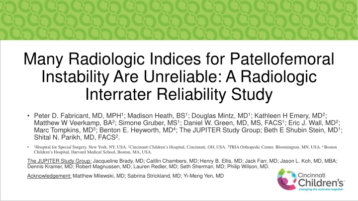
Reliability Study on Radiologic Indices for Patellofemoral Instability
Radiologic indices play a crucial role in the assessment and treatment planning for patellofemoral instability in children and adolescents. This study evaluates the interrater reliability of common radiographic parameters used to assess anatomic risk factors and aims to enhance the reliability of measurements with poor interrater agreement.

Download Presentation

Please find below an Image/Link to download the presentation.
The content on the website is provided AS IS for your information and personal use only. It may not be sold, licensed, or shared on other websites without obtaining consent from the author.If you encounter any issues during the download, it is possible that the publisher has removed the file from their server.
You are allowed to download the files provided on this website for personal or commercial use, subject to the condition that they are used lawfully. All files are the property of their respective owners.
The content on the website is provided AS IS for your information and personal use only. It may not be sold, licensed, or shared on other websites without obtaining consent from the author.
E N D
Presentation Transcript
Many Radiologic Indices for Patellofemoral Instability Are Unreliable: A Radiologic Interrater Reliability Study Peter D. Fabricant, MD, MPH1; Madison Heath, BS1; Douglas Mintz, MD1; Kathleen H Emery, MD2; Matthew W Veerkamp, BA2; Simone Gruber, MS1; Daniel W. Green, MD, MS, FACS1; Eric J. Wall, MD2; Marc Tompkins, MD3; Benton E. Heyworth, MD4; The JUPITER Study Group; Beth E Shubin Stein, MD1; Shital N. Parikh, MD, FACS2. 1Hospital for Special Surgery, New York, NY, USA. 2Cincinnati Children s Hospital, Cincinnati, OH, USA.3TRIA Orthopedic Center, Bloomington, MN, USA.4 Boston Children s Hospital, Harvard Medical School, Boston, MA, USA. The JUPITER Study Group: Jacqueline Brady, MD; Caitlin Chambers, MD;Henry B. Ellis, MD; Jack Farr, MD; Jason L. Koh, MD, MBA; Dennis Kramer, MD; Robert Magnussen, MD; Lauren Redler, MD; Seth Sherman, MD; Philip Wilson, MD. Acknowledgement: Matthew Milewski, MD; Sabrina Strickland, MD; Yi-Meng Yen, MD
Disclosure The authors have nothing to disclose
Introduction Surgical decision making for children and adolescents with patellofemoral instability relies heavily on accurate and consistent assessment of anatomic risk factors The assessment of risk factors from radiographs and MRIs can help predict risk of recurrence, guide treatment plan and dictate surgical procedures
Purpose 1. To evaluate the interrater reliability of common radiographic parameters used to evaluate anatomic risk factors for patellofemoral instability 2. To attempt to improve reliability for measurements that demonstrate unacceptable interrater reliability.
Methods Reliability study on prospectively collected data 2 fellowship-trained musculoskeletal radiologist assessed 50 patients with patellofemoral instability. 22 M / 28 F Average age: 14 yrs (+/-2) 27 measurements were performed on X-rays and MRI including: Trochlear dysplasia Caton-Deschamps index Patellar tilt
Variables View Units/Categorical Choices Lateral x-ray Lateral x-ray Lateral x-ray Lateral x-ray Axial x-ray Present or Absent Millimeters Present or Absent Number without units Present or Absent Trochlea Crossing Sign Trochlear Bump Double Contour Sign Caton-Deschamps Index Medial Patella Avulsion Fracture Axial x-ray Axial x-ray Degrees Lateral patellofemoral angle Lateral patellofemoral tilt Angle opens laterally (no tilt), Angle opens medially (tilt present), Lines are parallel (tilt present) Degrees None, Stage 1, Stage 2, Stage 3, Stage 4 Axial x-ray Axial x-ray Congruence angle Patellofemoral arthritis (Iwano Classification) Kellgren Lawrence: medial Kellgren Lawrence: lateral Leg Length Inequality (only skeletally immature patients) AP View AP View Grade 1, Grade 2, Grade 3, Grade 4 Grade 1, Grade 2, Grade 3, Grade 4 No difference, Right > Left, Left > Right Millimeters Standing AP, hips to ankles Standing AP, hips to ankles Leg length Difference (only skeletally immature patients) Mechanical axis: left Standing AP, hips to ankles Varus 1, Varus 2, Varus 3, Neutral, Valgus 1, Valgus 2, Valgus 3 Standing AP, hips to ankles Varus 1, Varus 2, Varus 3, Neutral, Valgus 1, Valgus 2, Valgus 3 Mechanical axis: right MRI MRI Present or Absent Effusion Bone edema None, Lateral femoral condyle, medial patellar facet, multiple location MRI MRI MRI Millimeters Trochlear bump Location of MPFL Injury Tibial tubercle to trochlear groove distance (TTTG) none, patella, femur, mid, combined Millimeters MRI MRI Degrees Sulcus angle Trochlear dysplasia: Dejour classification Type A, Type B, Type C, Type D MRI MRI MRI MRI MRI Degrees Millimeters Present or Absent None, patella, trochlea, both, other Millimeters Patellar tilt Patellar subluxation distance Osteochondral fracture Cartilage injury Trochlear Depth
Methods Intraclass correlation coefficients (ICC) were calculated for each measurement For all measurements with unacceptable reliability (ICC < 0.6), authors discussed consensus methods to improve reliability. Raters then re-measured a subset of 20 new patients. All measurements with an ICC less than 0.6 after consensus building and re-measurement were considered to be unreliable.
Results First round interrater reliability was acceptable for 5 radiographic measurements and 7 MRI measurements. After consensus building 4 additional radiographic measurements and 2 MRI measurements were reliable. 6 radiographic measurements and 3 MRI measurements remained unacceptably unreliable even after consensus training.
First Assessment Second Assessment Reliability Reliable with Training N ICC N ICC Initially Reliable Unreliable Variables X-Ray Trochlea crossing sign Trochlear bump Double contour sign Caton-Deschamps Index Medial patella avulsion fracture Lateral patellofemoral angle Lateral patellofemoral tilt (leave as unreliable) 33 34 34 47 50 44 0.455 0.205 0.38 0.644 0.43 0.768 10 7 10 - 18 - 0.625 0.435 -0.2 - 0.779 - X X X X X X 50 0.256 18 0 X Congruence angle Patellofemoral arthritis Kellgren-Lawrence: medial Kellgren-Lawrence: lateral Leg length inequality Leg length difference Mechanical axis: left Mechanical axis: right MRI Effusion Bone bruise Location of MPFL injury TT-TG distance Trochlear bump Trochlear dysplasia: Dejour classification 44 50 50 50 35 15 49 50 -0.133 0.111 0 0 0.092 0.985 0.777 0.665 18 18 - - 17 - - - 0.768 -0.062 - - 0.925 - - - X X X X X X X X 46 45 43 44 20 0.866 0.961 0.639 0.706 0.861 - - - - - - - - - - X X X X X 42 0.174 19 0.211 X Sulcus angle Patellar tilt Patellar subluxation distance Osteochondral fracture Cartilage injury Trochlear Depth 11 44 44 42 38 20 0.339 0.841 0.552 0.322 0.245 0.743 20 - 19 19 20 - 0.684 - 0.561 -0.108 0.686 - X X X X X X
Results Radiographs were reliable for Caton Deschamps Index and leg length discrepancy / alignment MRI were reliable for trochlear dysplasia and patellar tilt Dejour classification for trochlear dysplasia was unreliable even after consensus training
Conclusion This study found that many of the clinically relevant patellofemoral instability measurements have unacceptably low reliability across raters. However, with additional training and better definitions, some measurements do improve across raters. There are several measurements that did not improve even after additional consensus building and training. This may be due to ambiguity, the difficultly of the measurement, or for indices that are rarely used. Physicians should ensure that they are using a standardized definition or technique for radiographic measurements in patients with patellofemoral instability. Unreliable measurements should not be used in clinical decision making process.
References 1. Dejour H, Walch G, Nove-Josserand L, Guier C. Factors of patellar instability: An anatomic radiographic study. Knee Surgery, Sport Traumatol Arthrosc. 1994;2(1):19-26. doi:10.1007/BF01552649. 2. Smith TO, Davies L, Toms AP, Hing CB, Donell ST. The reliability and validity of radiological assessment for patellar instability. A systematic review and meta-analysis. Skeletal Radiol. 2011;40(4):399-414. 3. Lippacher S, Dejour D, Elsharkawi M, et al. Observer Agreement on the Dejour Trochlear Dysplasia Classification: A Comparison of True Lateral Radiographs and Axial Magnetic Resonance Images. Am J Sports Med. 2012;40(4):837-843. doi:10.1177/0363546511433028. 4. Pfirrmann A, Zanetti M, Romero J, Hodler J. Femoral Trochlear Dysplasia: MR Findings. Radiology. 2000;216:858-864. 5. Ye Q, Yu T, Wu Y, Ding X, Gong X. Patellar instability: The reliability of magnetic resonance imaging measurement parameters. BMC Musculoskelet Disord. 2019;20(1):317. doi:10.1186/s12891-019-2697-7.