Renal Tubule Function
The renal tubule is a complex structure responsible for reabsorption and secretion processes essential for maintaining electrolyte balance and fluid volume in the body. Thiazide diuretics, loop diuretics, and K+-sparing diuretics target specific transport mechanisms within the nephron to alter electrolyte handling. Learn about the different segments of the renal tubule and the mechanisms of action of various diuretics.

Download Presentation

Please find below an Image/Link to download the presentation.
The content on the website is provided AS IS for your information and personal use only. It may not be sold, licensed, or shared on other websites without obtaining consent from the author.If you encounter any issues during the download, it is possible that the publisher has removed the file from their server.
You are allowed to download the files provided on this website for personal or commercial use, subject to the condition that they are used lawfully. All files are the property of their respective owners.
The content on the website is provided AS IS for your information and personal use only. It may not be sold, licensed, or shared on other websites without obtaining consent from the author.
E N D
Presentation Transcript
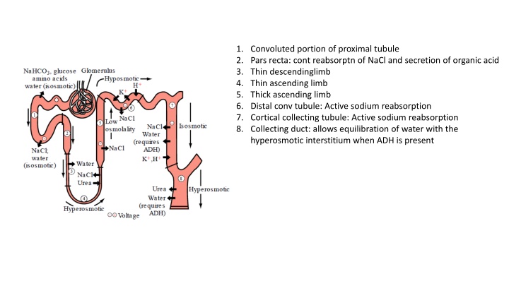
1. Convoluted portion of proximal tubule 2. Pars recta: cont reabsorptn of NaCl and secretion of organic acid 3. Thin descendinglimb 4. Thin ascending limb 5. Thick ascending limb 6. Distal conv tubule: Active sodium reabsorption 7. Cortical collecting tubule: Active sodium reabsorption 8. Collecting duct: allows equilibration of water with the hyperosmotic interstitium when ADH is present
INHIBITORS OF NA+-CL- SYMPORT (THIAZIDE AND THIAZIDELIKE DIURETICS) inhibitors of Na+-Cl- symport increase Na+ and Cl- excretion. However, thiazides are only moderately efficacious (i.e., maximum excretion of filtered load of Na+ is only 5%) because approximately 90% of the filtered Na+ load is reabsorbed before reaching the DCT. Some thiazide diuretics also are weak inhibitors of carbonic anhydrase, an effect that increases HCO3- and phosphate excretion and probably accounts for their weak proximal tubular effects. Like inhibitors of Na+-K+-2Cl- symport, inhibitors of Na+-Cl- symport increase the excretion of K+ and titratable acid by the same mechanisms discussed for loop diuresis.
INHIBITORS OF NA+-K+-2CL- SYMPORT (LOOP DIURETICS, HIGH-CEILING DIURETICS All inhibitors of Na+-K+-2Cl- symport increase the urinary excretion of K+ and titratable acid. This effect is due in part to increased delivery of Na+ to the distal tubule. The mechanism by which increased distal delivery of Na+ enhances excretion of K+ and H+ is discussed in the section on inhibitors of Na+ channels. Other mechanisms contributing to enhanced K+ and H+ excretion include flow-dependent enhancement of ion secretion by the collecting duct, nonosmotic vasopressin release, and activation of the renin-angiotensin-aldosterone axis
INHIBITORS OF RENAL EPITHELIAL NA+ CHANNELS (K+-SPARING DIURETICS) Carbonic anhydrase inhibitors, loop diuretics, and thiazide diuretics increase the delivery of Na+ to the late distal tubule and collecting duct, a situation that often is associated with increased K+ and H+ excretion. It is likely that the elevation in luminal Na+ concentration in the distal nephron induced by such diuretics augments depolarization of the luminal membrane and thereby enhances the lumen-negative VT, which facilitates K+ excretion. increased distal delivery of Na+ is not the only mechanism by which diuretics increase K+ and H+ excretion. Activation of the renin-angiotensin-aldosterone axis by diuretics also contributes to diuretic-induced K+ and H+ excretion, As the sodium rushes back into the cell the positive sodium ions raise the charge inside the cell from negative to positive. Once the interior of the cell becomes positively charged, depolarization of the cell is complete
Effects of aldosterone on late distal tubule and collecting duct and diuretic mechanism of aldosterone antagonists. Epithelial cells in the late distal tubule and collecting duct contain cytosolic MRs that have a high affinity for aldosterone AIP, aldosterone-induced proteins; ALDO, aldosterone; MR, mineralocorticoid receptor; CH, ion channel 1, activation of membrane-bound Na+ channels 2, redistribution of Na+ channels from cytosol to membrane; 3, de novo synthesis of Na+ channels; 4, activation of membrane- bound Na+, K+-ATPase; 5, redistribution of Na+,K+-ATPase from cytosol to membrane; 6, de novo synthesis of Na+,K+-ATPase; 7, changes in permeability of tight junctions; 8, increased mitochondrial production of ATP
Mechanism of action of aldosterone Epithelial cells in the late distal tubule and collecting duct contain cytosolic MRs that have a high affinity for aldosterone Aldosterone enters the epithelial cell from the basolateral membrane and binds to MRs; the MR-aldosterone complex translocates to the nucleus, where it binds to specific sequences of DNA (hormone-responsive elements) and thereby regulates the expression of multiple gene products called aldosterone-induced proteins (AIPs). Figure illustrates some of the proposed effects of AIPs, including activation of "silent" Na+ channels and "silent" Na+ pumps that pre-exist in the cell membrane, alterations in the cycling of Na+ channels and Na+ pumps between the cytosol and cell membrane such that more channels and pumps are located in the membrane, increased expression of Na+ channels and Na+ pumps, changes in permeability of the tight junctions, and increased activity of enzymes in the mitochondria that are involved in ATP production. The precise mechanisms by which AIPs alter transport are incompletely understood. However, the net effect of AIPs is to increase Na+ conductance of the luminal membrane and sodium pump activity of the basolateral membrane. Consequently, transepithelial NaCl transport is enhanced, and the lumen-negative transepithelial voltage is increased. The latter effect increases the driving force for secretion of K+ and H+ into the tubular lumen.
Electrolyte and Water Composition of Body Fluid Compartments Components Plasma Interstitial fluid Intracellular fluid Volume, H2O (TBW = 42 L) 3.5 L 10.5 L 28 L Na+ 142 145 12 K+ 4 4 156 2.4 2-3 2.3 Ca+2 Mg2+ 2 1-2 26 Trace elements 1 - - Total cations 155 Cl 103 114 4 HCO 27 31 12 Protein 16 - 55 Organic acids 5 HPO2 2 SO2 1 Total anions 154
Reference interval of Sodium: Urinary sodium excretion = 120-240 mmol/day with large diurnal variation At night = 20% of the peak 136-145 mmol/L (Adult) 128-148 mmol/L (New born at 48 h) Approx 127 mmol/L (From Umbilical cord) Hyponatremia typically manifests clinically as (1) nausea, (2) generalize weakness, and (3) mental confusion. <120 mmol/L: mental confusion <110 mmol/L : Ocular palsy 90-105 mmol/L: Severe mental impairment
Hypernatremia Plasma sodium > 150 mmol/L Symptoms are primarily neurologic (because of neuronal cell loss of H2O into the ECF) 1.Tremors 2.Irritability 3.Ataxia 4.Confusion 5.coma
HYPOKALEMIA 1.Muscle weakness 2.Irritability 3.Paralysis 4.Tachycardia 5.Cardiac conduction defect 6.Flattened T wave 7.Cardiac arrest Reference interval of K+: Serum=3.5-5.0 mmol/L (Adult) Plasma= 3.4-4.8 mmol/L (Adult) 3.7-5.9 mmol/L ( Newborn) CSF= 70% that of plasma
Hypokalemia (continued) Metabolic Alkalosis
HYPERKALEMIA 1. Mental confusion 2. Weakness 3. Tingling 4. Flaccid paralysis of the extremities 5. Weakness of the respiratory muscles 6. Bradicardia 7. Conduction defects 8. Peripheral vascular collapse : Prolonged severe hyperkalemia >7 mmol/L 9. Cardiac arrest


")