Reproducibility of Diagnosis Assessment in ELN Low-Risk Registry
This research aims to assess the reproducibility of diagnosis in low-risk ELN registry cases by reviewing selected slides from various countries. The methodology involves a panel of experts and trainee physicians, with results compared to original findings. Detailed morphologic characteristics are examined, with discrepancies calculated using a test image. Essential data sets are prepared for review, focusing on key parameters for accurate diagnosis.

Download Presentation

Please find below an Image/Link to download the presentation.
The content on the website is provided AS IS for your information and personal use only. It may not be sold, licensed, or shared on other websites without obtaining consent from the author.If you encounter any issues during the download, it is possible that the publisher has removed the file from their server.
You are allowed to download the files provided on this website for personal or commercial use, subject to the condition that they are used lawfully. All files are the property of their respective owners.
The content on the website is provided AS IS for your information and personal use only. It may not be sold, licensed, or shared on other websites without obtaining consent from the author.
E N D
Presentation Transcript
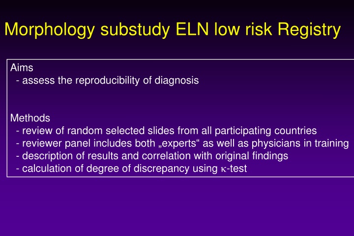
Morphology substudy ELN low risk Registry Aims - assess the reproducibility of diagnosis Methods - review of random selected slides from all participating countries - reviewer panel includes both experts as well as physicians in training - description of results and correlation with original findings - calculation of degree of discrepancy using -test
Methods in detail - Random selection of a total of 100 cases from all countries taking into account the amount of included patients (for example more from France as compared to Germany -> already done by Alex Smith -2 marrow slides (preferably also iron staining) and 1 blood film from each selected case should be sent to D sseldorf, where the morphology review will take place -> action by country coordinators
Methods in detail Preparation of minimal required data set of selected cases necessary for review, to be sent to D sseldorf: - ID, (Country, Hospital: only available to Alex Smith) - age at diagnosis - Hb, WBC, Platelets, Defferential count, LDH, Bilirubin, Coombs test - cytogenetic finding at diagnosis: necessary for WHO classification - no original morphology report -> to be done by Alex Smith
Morphology assessment in detail Description of Morphologic characeristics, to be filled in a file during the morphology meeting I) Peripheral blood - % of blasts - % of monocytes - signs of Dysplasia - Anisometry of platelts? - Macroplatelets? - Pseudopelger? - Hypogranulation? - cytoplasma anomalies of erythrocytes?
Morphology assessment in detail Description of Morphologic characeristics, to be filled in a file during the morphology meeting II) Marrow 1) Erythropoiesis - % of erythropoiesis - Clear signs of dysplasia? - Nuclear abnormalities? - Cytoplasma abnormalities? - Ring sideroblasts? (%) 2) Megakaryopoiesis - Assessment of 25 megakaryocytes - Clear signs of dysplasia? (>10%) - Micromegakaryocytes ? - Mononuclear megakaryocytes? - Megas with spreaded nuclei?
Morphology assessment in detail Description of Morphologic characeristics 3) Granulopoiesis - Clear signs of dysplasia? (>10%) - Pseudopelger? - Hypogranulation? - % Medullary blast (if Erythropoiesis >50%, count blasts in the non-erythropoietic cells) 4) Other items - Cellularity (Hypo vs. Normo vs. Hyper) - Plasma cells increased? - Lymphoid cells increased? - Iron load? - Other stainings available?
Morphology assessment in detail Diagnostic categories RCUD (bicytopenia) RC RA RN MDS unclass RCUD with pancytopenia RCUD/RCMD with blasts in blood RCUD without dysplasia but with clonal marker RCMD MDS with del(5q) RARS-T RAEB I RAEB II, RAEB-T, AML, CMML I, CMMLII, ICUS No MDS at all
Methods to analyse the results 1) Within the panel - Discussion of cases during meeting - Report on interobserver variation within experts - Report on possible discrepancies between experts and non-experts - description of staining techniques and quality of slides - comparison of methods to read the slides - correlation of WHO types with hematologic and cytogenetic findings 2) Panel vs. Original reports - Send list of WHO results of the morphology panel to Alex Smith - Comparison of original reports with the results of the panel by k-test (and/or other tests to be done my Alex Smith) to describe amount of discrepancies 3) Final report on - Validation and reliability of the cases included into the ELN low risk R
Methods to analyse the results 3) Final report on - Validation and reliability of the cases included into the ELN low risk Registry to be done within the panel and Alex Smith
Morphology Panel Chairs: Proposed Experts Argiris Symenoidis, Greece Jaroslaw Cermak, Czech Rep. Anna Porwit, Sweden Teresa Valespie?, Spain Proposed non-experts Louise deSwart, Netherlands Judith Neukirchen, Germany By David Bowen, UK Others Ulrich Germing and Marius MacKenzie